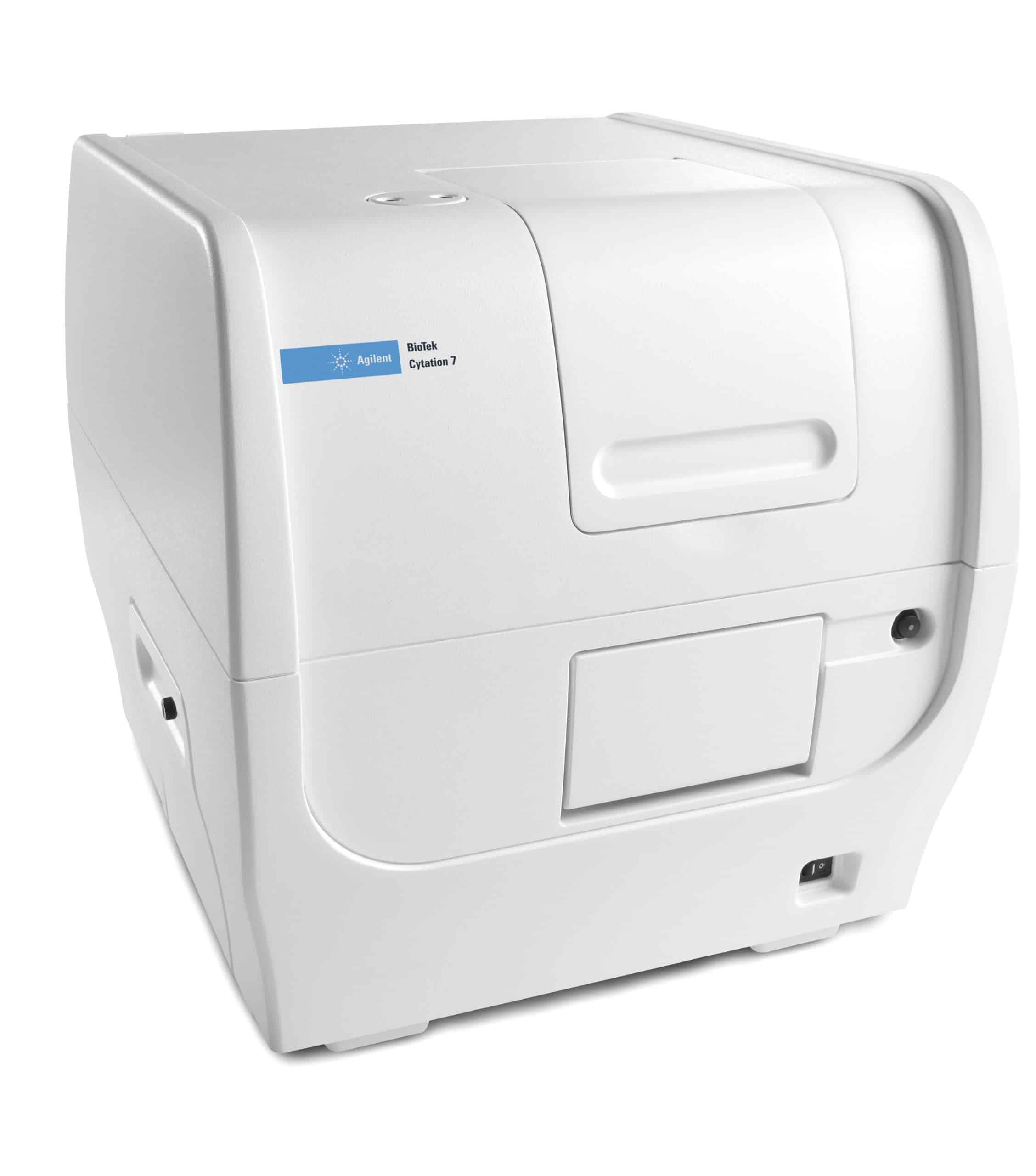
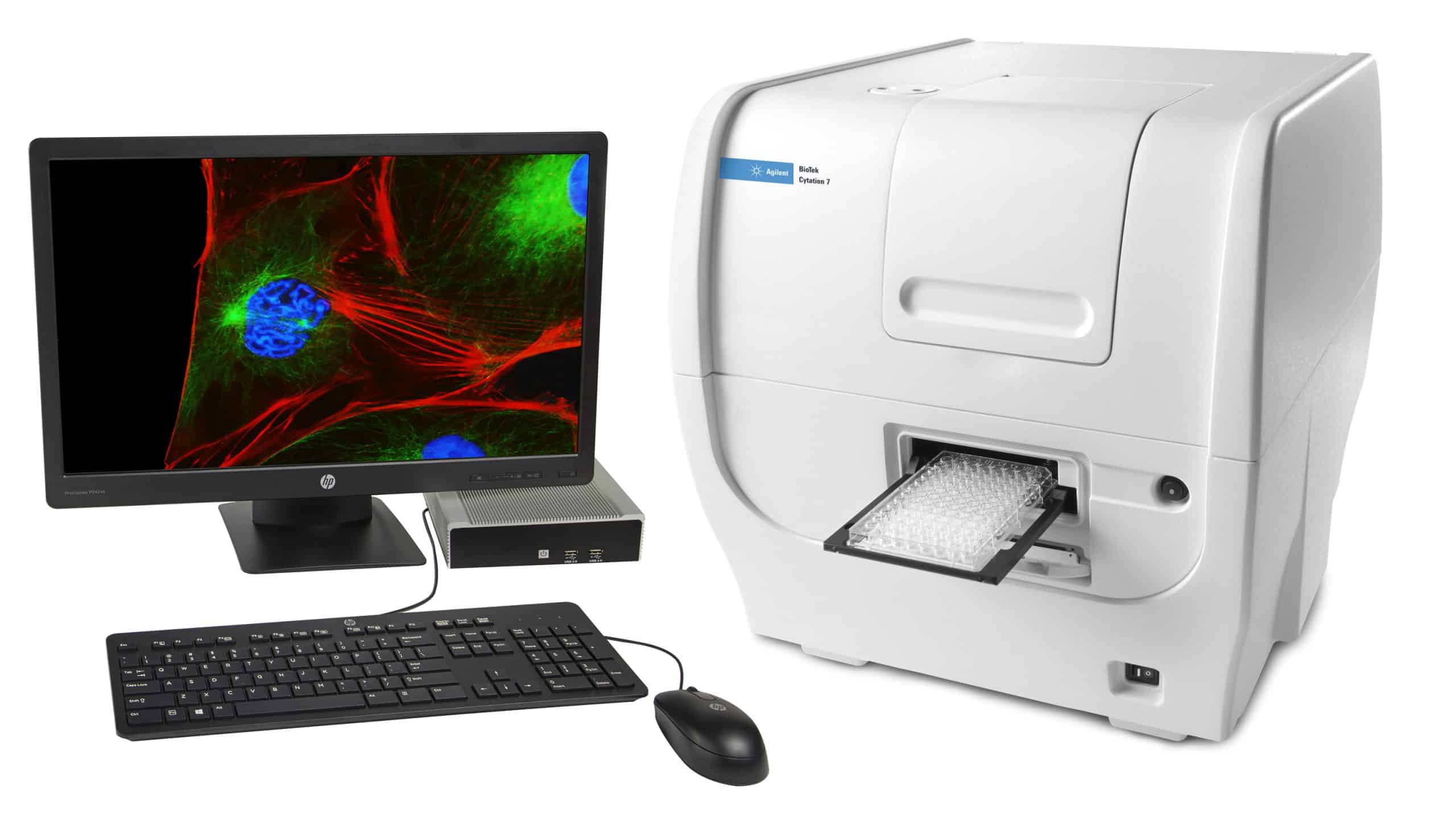
BioTek Cytation 7 cell imaging multimode reader combines automated digital upright and inverted widefield microscopy with conventional multimode microplate reading. Cytation 7 includes continually variable bandpass monochromators for versatility and performance for general multimode plate reader applications.
The inverted microscopy module provides sample visualization and the upright microscopy module with reflected light imaging enables even more applications, including ELISpot and fast slide scanning. Cytation 7 is controlled by Gen5 software, which combines ease-of-use with powerful processing and analysis capabilities.
| Cytation 7 | Cytation 5 | Cytation 1 | |
| General | |||
| Detection modes | UV-Vis absorbance | UV-Vis absorbance | UV-Vis absorbance |
| Fluorescence intensity | Fluorescence intensity | Fluorescence intensity | |
| Luminescence | Luminescence | Luminescence | |
| Fluorescence polarization | Fluorescence polarization | ||
| Time-resolved fluorescence | Time-resolved fluorescence | ||
| Alpha | |||
| Read methods | Endpoint, kinetic, spectral scanning, well area scanning | Endpoint, kinetic, spectral scanning, well area scanning | Endpoint, kinetic, spectral scanning, well area scanning |
| Microplate types | Monochromator: 6- to 384-well plates | Monochromator: 6- to 384-well plates | Monochromator: 6- to 384-well plates |
| Imaging: 6- to 1536-well plates | Filters: 6- to 1536-well plates | Filters: 6- to 1536-well plates | |
| Imaging: 6- to 1536-well plates | Imaging: 6- to 1536-well plates | ||
| Other labware supported | Microscope slides, Petri and cell culture dishes, cell culture flasks (T25), counting chambers (hemocytometer) | Microscope slides, Petri and cell culture dishes, cell culture flasks (T25), counting chambers (hemocytometer) | Microscope slides, Petri and cell culture dishes, cell culture flasks (T25), counting chambers (hemocytometer) |
| Take3 Micro-Volume Plates | Take3 Micro-Volume Plates | Take3 Micro-Volume Plates | |
| Temperature control | 4-Zone incubation to 45 °C with Condensation Control; | 4-Zone incubation to 65 °C with Condensation Control; | 4-Zone incubation to 45 °C with Condensation Control™; |
| +0.5 °C at 37 °C | +0.2 °C at 37 °C | Variation +0.2 °C at 37 °C | |
| Cooling | Optional Peltier Cooling Module maintains internal temperature with < 1 °C rise over ambient. Provides internal cooling after incubated processes. | Optional Peltier Cooling Module maintains internal temperature with < 1 °C rise over ambient. Provides internal cooling after incubated processes. | Optional Peltier Cooling Module maintains internal temperature with < 1 °C rise over ambient. Provides internal cooling after incubated processes. |
| Shaking | Linear, orbital, double orbital | Linear, orbital, double orbital | Linear, orbital, double orbital |
| Software | Gen5™ Microplate Reader and Imager Software included | Gen5™ Microplate Reader and Imager Software | Gen5™ Microplate Reader and Imager Software |
| Gen5 Secure for 21 CFR Part 11 compliance (option) | Gen5 Image+ and Image Prime software available for full image analysis | Gen5 Image+ and Image Prime software available for full image analysis | |
| Gen5 Secure available for 21 CFR Part 11 | |||
| Gen5 Secure for 21 CFR Part 11 compliance (option) | |||
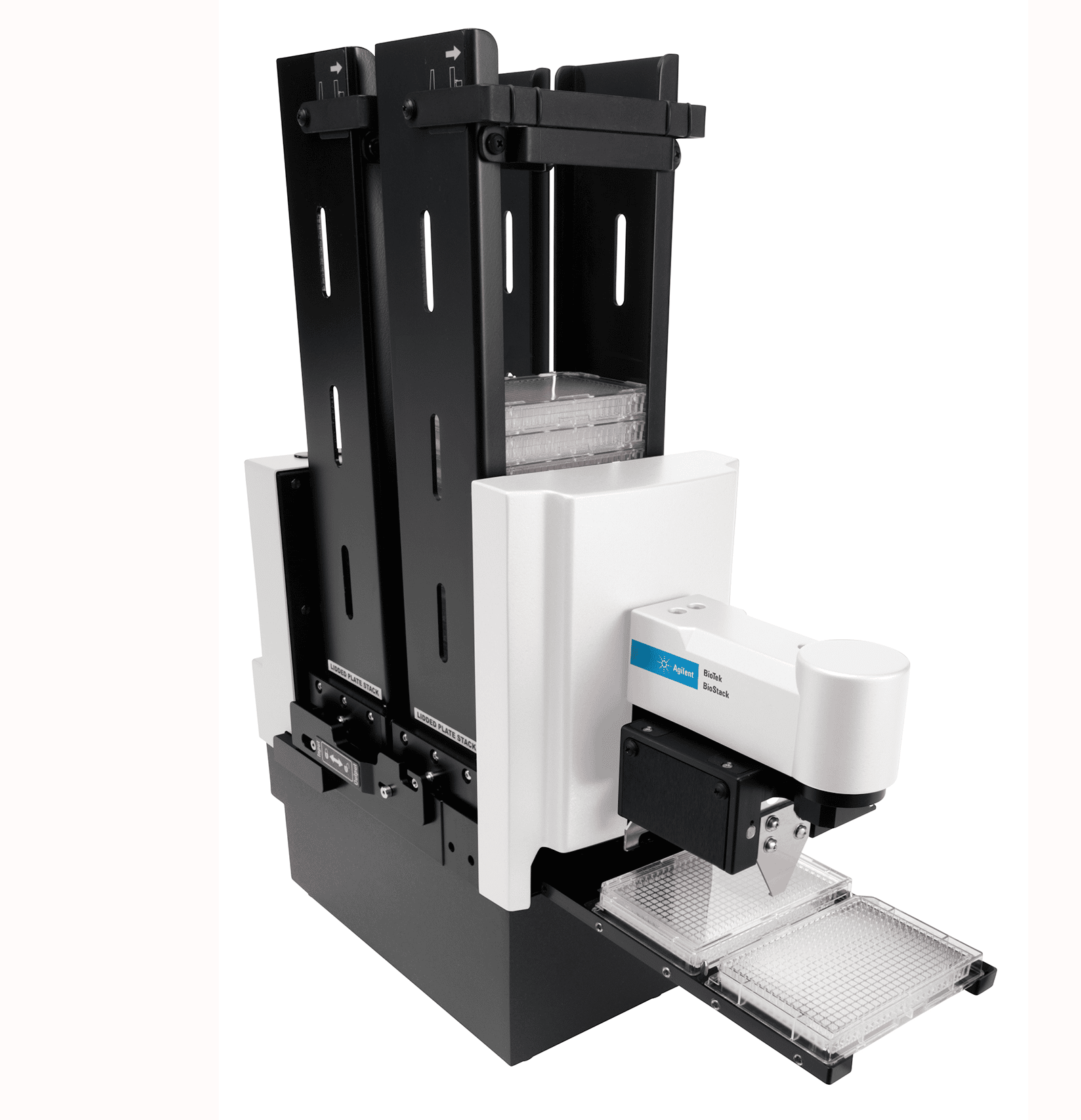
| Automation | BioStack and 3rd party automation compatible | BioStack and 3rd party automation compatible | BioStack and 3rd party automation compatible |
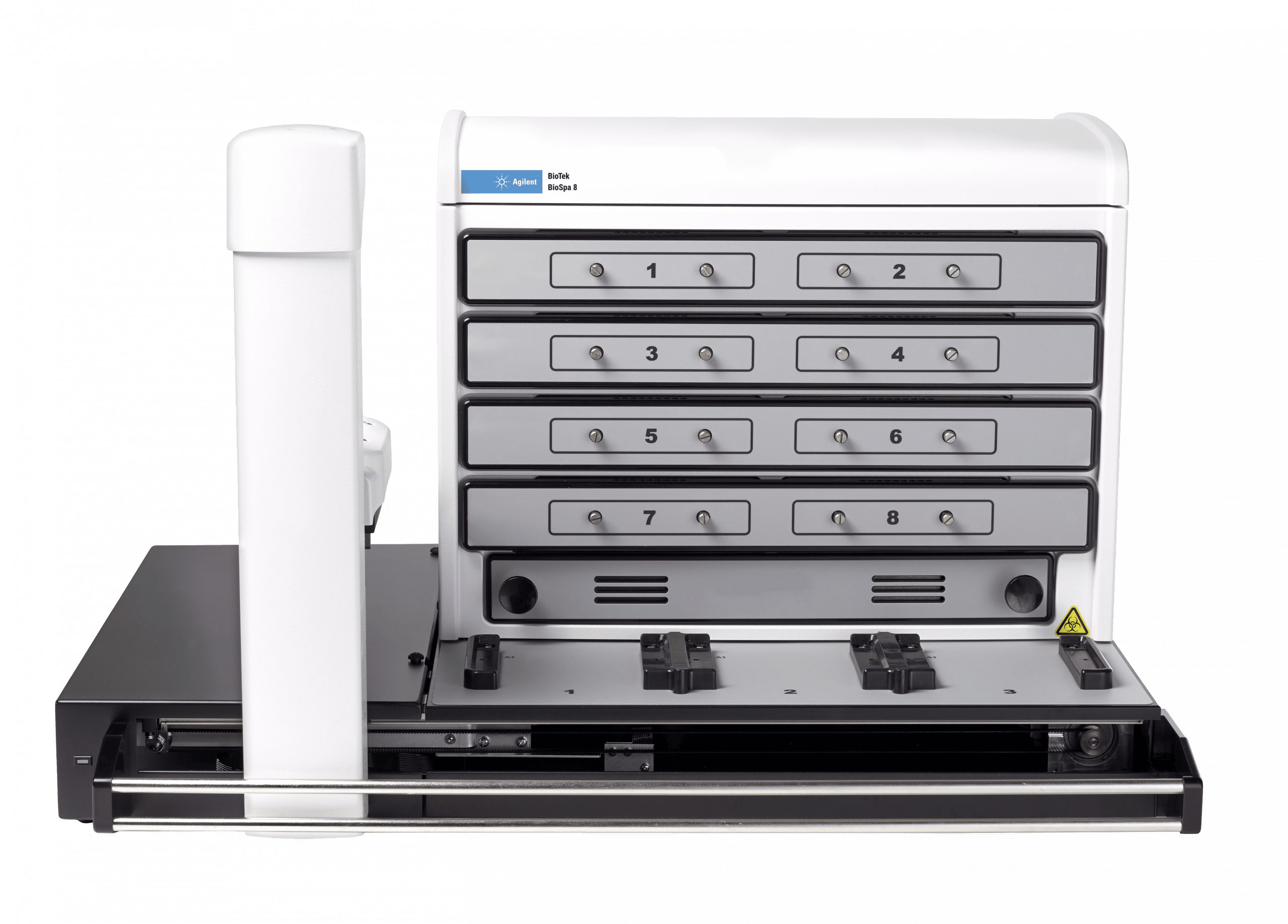
| BioSpa 8 Automated Incubator compatible | BioSpa 8 Automated Incubator compatible | BioSpa 8 Automated Incubator compatible | |
| CO2 and O2 control (option) | Range: 0 – 20% (CO2); 1 – 19% (O2) | Range: 0 – 20% (CO2); 1 – 19% (O2) | 0 – 20% CO2 control and 1 – 19% O2 control, with optional Gas Controller |
| Models for both CO2 and O2 or CO2 only are available | Models for both CO2 and O2 or CO2 only are available | Models for both CO2 and O2 or CO2 only are available | |
| Imaging – Inverted Microscope | |||
| Imaging mode | Fluorescence, color brightfield, user-selectable brightfield/high contrast brightfield | Fluorescence, brightfield, high contrast brightfield, color brightfield and phase contrast | Fluorescence and high contrast brightfield (2.5x Zeiss, 4x, 10x, 20x, 40x and 60x) |
| Imaging method | Single color, multi-color, time lapse, montage, z-stacking, z-stack montage | Single color, multi-color, montage, time lapse, z-stacking | Single color, multi-color, montage, time lapse, z-stacking |
| Image processing | Z-projection, digital phase contrast, stitching | Z-projection, digital phase contrast, stitching | Z-projection, image stitching |
| Camera | 16-bit Sony CMOS wide field of view monochrome camera | 16-bit Sony CMOS, standard or wide FOV | 16-bit gray scale, Sony |
| Objective capacity | 6-position automated turret for user-replaceable objectives | 6-position automated turret for user-replaceable objectives | 2 user-replaceable objectives |
| Objectives available | 1.25x, 2.5x (2.25x eff),2.5x (2.75x eff), 4x,10x, 20x, 40x, 60x | 1.25x, 2.5x (2.25x eff),2.5x (2.75x eff), 4x,10x, 20x, 40x, 60x | 1.25x, 2.5x (2.25x eff),2.5x (2.75x eff), 4x,10x, 20x, 40x, 60x |
| Phase objectives available | 4x, 10x, 20x, 40x | ||
| Image filter cube capacity | 4 user-replaceable fluorescence cubes, plus brightfield channel | 4 user-replaceable fluorescence cubes plus brightfield channel | 4 user-replaceable fluorescence cubes plus brightfield channel |
| Imaging filter cubes available | DAPI, CFP, GFP, YFP, RFP, Texas Red, CY5, CY7, Acridine Orange, CFP-FRET, CFP-YFP FRET, Chlorophyll, Phycoerythrin (PE), Propidium Iodide, CY5.5, TagBFP, Tag BFP-FRET, GFP (Ex)-CY5 (Em), RFP (Ex)-CY5 (Em), Alexa 568, Ex 377/Em 647, Oxidized roGFP2, TRITC | DAPI, CFP, GFP, YFP, RFP, Texas Red, CY5, CY7,Acridine Orange (ACR OR), CFP-YFP FRET, propidium,Iodide, chlorophyll, phycoerythrin, CY5.5, TagBFP, Alexa568, Ex377 / Em647 | DAPI, CFP, GFP, YFP, RFP, Texas Red, CY5, CY7,Acridine Orange (ACR OR), CFP-YFP FRET, propidium,Iodide, chlorophyll, phycoerythrin, CY5.5, TagBFP, Alexa568, Ex377 / Em647 |
| Imaging LED cubes available | 365 nm, 390 nm, 465 nm, 505 nm, 523 nm, 554 nm, 590 nm, 623 nm, 655 nm, 740 nm | 365 nm, 390 nm, 465 nm, 505 nm, 523 nm, 590 nm, 623 nm, 655 nm, 740 nm | 365 nm, 390 nm, 465 nm, 505 nm, 523 nm, 590 nm, 623 nm, 655 nm, 740 nm |
| Automated functions | Autofocus, user-trained autofocus, autoexposure, auto-LED intensity | Autofocus, auto LED intensity, auto exposure | Autofocus, auto LED intensity, auto exposure |
| Autofocus method | Image-based autofocus | Image-based autofocus | Image-based autofocus |
| User-trained autofocus | User-trained autofocus | User-trained autofocus | |
| Laser autofocus (option) | Laser autofocus (option) | Laser autofocus (option) | |
| Positional controls | Software Control | Software control | Software control |
| Joystick controller (option) | Joystick control (option) | ||
| Image collection rate | Image-based autofocus: | Image-based autofocus: | Image-based autofocus: |
| 96 wells, 1 color (DAPI), 4x, 6 minutes | 96 wells, 1 color (DAPI), 4x, 6 minutes | 96 wells, 1 color (DAPI), 4x, 6 minutes | |
| 96 wells, 3 colors, 4x, 12 minutes | 96 wells, 3 colors, 4x, 12 minutes | ||
| Laser autofocus: | |||
| Laser autofocus: | |||
| 96 wells, 1 color (DAPI), 4x, <3 minutes | Laser autofocus: | ||
| 96 wells, 1 color (DAPI), 4x, <3 minutes | |||
| 96 wells, 1 color (DAPI), 4x, <3 minutes | |||
| 96 wells, 3 colors, 4x, <7 minutes, 30 seconds | |||
| 96 wells, 3 colors, 4x, <7 minutes, 30 seconds | |||
| Burst Mode: 10 fps, single well, single color at <= 5Oms integration time | |||
| Burst Mode: 10 fps, single well, single color at <= 50ms integration time | |||
| Image Analysis Software option | Gen5 Image+: Image analysis | Gen5 Image+: Image analysis | Gen5 Image+: Image analysis |
| Gen5 Image Prime: Advanced image analysis | Gen5 Image Prime: Advanced image analysis | Gen5 Image Prime: Advanced image analysis | |
| Gen5 Secure: 21 CFR Part 11 compliant features | Gen5 Secure: 21 CFR Part 11 compliant features | Gen5 Secure: 21 CFR Part 11 compliant features | |
| Imaging – Upright Microscope | |||
| Imaging modes | Reflected color brightfield; transmitted color brightfield | ||
| Imaging methods | Single image, montage, time lapse, Z-stacking | ||
| Image processing | Z-projection, digital phase contrast, stitching | ||
| Camera | 16-bit Sony CMOS wide field of view color camera | ||
| Lenses | Finder scope, 2x, 4x, 8x | ||
| Positional controls | Software Control | ||
| Joystick controller (option) | |||
| Image collection rate | Entire 100 mm dish @ 1x: <2:40 minutes | ||
| Entire microscope slide @1x: <1:15 minutes | |||
| 96-well ELISpot plate@ 1x: <5 minutes | |||
| Image Analysis Software option | Gen5 Image+: Image analysis | ||
| Gen5 Image Prime: Advanced image analysis | |||
| Gen5 Secure: 21 CFR Part 11 compliant features | |||
| Fluorescence Intensity | |||
| Light source | Xenon flash | Xenon flash | Xenon flash |
| Detector | PMT (red-shifted PMT option) | PMT for monochromator system | PMT |
| PMT for filter system | |||
| Wavelength selection | Quad monochromators (top/bottom) | Quad monochromators (top/bottom) | Deep blocking band pass filters / dichroic mirrors |
| Filters (top) | |||
| Wavelength range | 250 – 700 (900 nm option) | Monochromators: 250 – 700 nm (900 nm option) | Filters: 200 – 700 nm (850 nm option) |
| Filters: 200 – 700 nm (850 nm option) | |||
| Monochromator bandwidth | Variable, from 9 nm to 50 nm in 1 nm increments | Variable, from 9 nm to 50 nm in 1 nm increments | |
| Dynamic range | 7 decades | 7 decades | 7 decades |
| Sensitivity | Fluorescein 2.5 pM (0.25 fmol/well, 384-well plate) – top | Filters: | Fluorescein: 0.25 pM (0.025 fmol/well, 384-well plate) |
| Fluorescein 4 pM (0.4 fmol/well, 384-well plate) – bottom | Fluorescein 0.25 pM (0.025 fmol/well, 384-well plate) | ||
| Quad Monochromator: | |||
| Fluorescein 2.5 pM (0.25 fmol/well, 384-well plate) – top | |||
| Fluorescein 4 pM (0.4 fmol/well, 384-well plate) – bottom | |||
| Reading speed (kinetic) | 96 wells: 11 seconds | 96 wells: 11 seconds | 96 wells: 11 seconds |
| 384 wells: 22 seconds | 384 wells: 22 seconds | 384 wells: 22 seconds | |
| Luminescence | |||
| Wavelength range | 300 – 700 nm | 300 – 700 nm | 300 – 700 nm |
| Dynamic range | >6 decades | >6 decades | >6 decades |
| Sensitivity | 20 amol ATP (flash) | Monos: 20 amol ATP (flash) | 10 amol ATP (flash) |
| Filters: 10 amol ATP (flash), 100 amol (glow) | 100 amol (glow) | ||
| Fluorescence Polarization | |||
| Light source | Xenon flash | Xenon flash | |
| Detector | PMT | PMT | |
| Wavelength selection | Filters | Filters | |
| Wavelength range | 280 – 700 nm (850 nm option) | 400 nm – 700 nm | |
| Sensitivity | 1.2 mP standard deviation at 1 nM fluorescein | 1.2 mP standard deviation at 1 nM fluorescein | |
| Time-Resolved Fluorescence | |||
| Light source | Xenon flash | Xenon flash | |
| Detector | PMT | PMT | |
| Wavelength selection | Quad monochromators (secondary mode) | Filters | |
| Filters (top) | |||
| Wavelength range | Filters: 200 – 700 nm (850 nm option) | ||
| Sensitivity | Filters: Europium 40 fM (4 amol/well, 384-well plate) | Europium 40 fM (4 amol/well, 384-well plate) | |
| Monos: Europium 1200 fM (120 amol/well, 384-well plate) | |||
| Absorbance | |||
| Light source | Xenon flash | Xenon flash | Xenon flash |
| Detector | Photodiode | photodiode | photodiode |
| Wavelength selection | Monochromator | Monochromator | Monochromator |
| Wavelength range | 230 – 999 nm, 1 nm increment | 230 – 999 nm, 1 nm increment | 200 – 999 nm, 1 nm increment |
| Monochromator bandwidth | 4 nm (230 – 285 nm), 8 nm (>285 nm) | 4 nm (230 – 285 nm), 8 nm (>285 nm) | 2.4 nm |
| Dynamic range | 0 – 4.0 OD | 0 – 4.0 OD | 0 – 4.0 OD |
| Resolution | 0.0001 OD | 0.0001 OD | 0.0001 OD |
| Pathlength correction | Yes | yes | yes |
| Monochromator wavelength accuracy | +2 nm | +2 nm | + 2 nm |
| Monochromator wavelength repeatability | +0.2 nm | +0.2 nm | + 0.2 nm |
| OD accuracy | <1% at 3.0 OD | <1% at 2.0 OD | <1% at 2.0 OD |
| <3% at 3.0 OD | <3% at 3.0 OD | ||
| OD linearity | <1% from 0 to 3.0 OD | <1% from 0 to 3.0 OD | <1% from 0 to 3.0 OD |
| OD repeatability | <0.5% at 2.0 OD | <0.5% at 2.0 OD | <0.5% at 2.0 OD |
| Stray light | 0.03% at 230 nm | 0.03% at 230 nm | 0.03% at 230 nm |
| Reading speed (kinetic) | 96 wells: 11 seconds | 96 wells: 11 seconds | 96 wells: 11 seconds |
| 384 wells: 22 seconds | 384 wells: 22 seconds | 384 wells: 22 seconds | |
| Alpha Detection | |||
| Light source | 100 mW 680 nm laser | ||
| Detector | PMT | ||
| Wavelength selection | Filters (top) | ||
| Sensitivity | 100 amol LCK peptide (384-well low volume plate) | ||
| Reagent Injectors (option) | |||
| Supported detection modes | All modes | All modes | |
| Number | 2 syringe pumps | 2 syringe pumps | 2 syringe pumps |
| Supported labware | 6- to 384-well plates, Petri and cell culture dishes | 6- to 384-well plates, Petri and cell culture dishes | 6- to 384-well plates, Petri and cell culture dishes |
| Dead volume | 1.1 mL with back flush | 1.1 mL with back flush | 1.1 mL with back flush |
| Dispense volume | 5 – 1000 µL in 1 µL increments | 5 – 1000 µL in 1 µL increments | 5 – 1000 µL in 1 µL increments |
| Plate geometry | 6- to 384-well microplates | 6- to 384-well microplates | 6- to 384-well microplates |
| Dispense accuracy | +1 µL or 2% | +1 µL or 2% | +1 µL or 2% |
| Dispense precision | <2% at 50 – 200 µL | <2% at 50 – 200 µL | <2% at 50 – 200 µL |
| Physical Characteristics | |||
| Power | 150 Watts maximum consumption | 250 Watts maximum consumption. | 100-240 VAC, 50/60 Hz (24VDC external power supply, 150W min) |
| Dimensions | 20.2″ D x 16.4″ W x 17.5″ H | 20.2″ D x 16.4″ W x 17.5″ H | 20.2″ D x 16.4″ W x 17.5″ H |
| (51.4 cm D x 41.6 cm W x 44.5 cm H) | (51.4 cm D x 41.6 cm W x 44.5 cm H) | (51.4 cm D x 41.6 cm W x 44.5 cm H) | |
| Weight | 80 lbs (36.3 kg) | 80 lbs (36.3 kg) | 65 lbs (29 Kg) |
| Regulatory | |||
| Regulatory | CE and TUV marked. RoHS Compliant. Models for In Vitro Diagnostic use are available. | CE and TUV marked. RoHS Compliant. Models for In Vitro | CE and TUV marked. Models for In Vitro Diagnostic use are available. |
| Diagnostic use are available. | |||
| Specifications are subject to change. | |||
| GENERAL | |
| Detection modes | UV-Vis absorbance |
| Fluorescence intensity | |
| Luminescence | |
| Read methods | Endpoint, kinetic, spectral scanning, well area scanning |
| Microplate types | Monochromator: 6- to 384-well plates |
| Imaging: 6- to 1536-well plates | |
| Other labware supported | Microscope slides, Petri and cell culture dishes, cell culture flasks (T25), counting chambers (hemocytometer) |
| Take3 Micro-Volume Plates | |
| Temperature control | 4-Zone incubation to 45 °C with Condensation Control; +0.5 °C at 37 °C |
| Cooling | Optional Peltier Cooling Module maintains internal temperature with < 1 °C rise over ambient. Provides internal cooling after incubated processes. |
| Shaking | Linear, orbital, double orbital |
| Software | Gen5™ Microplate Reader and Imager Software included |
| Gen5 Secure for 21 CFR Part 11 compliance (option) | |
| Automation | BioStack and 3rd party automation compatible |
| BioSpa 8 Automated Incubator compatible | |
| CO2 and O2 control (option) | Range: 0 – 20% (CO2); 1 – 19% (O2) |
| Models for both CO2 and O2 or CO2 only are available | |
| IMAGING – INVERTED MICROSCOPE | |
| Imaging mode | Fluorescence, color brightfield, user-selectable brightfield/high contrast brightfield |
| Imaging method | Single color, multi-color, time lapse, montage, z-stacking, z-stack montage |
| Image processing | Z-projection, digital phase contrast, stitching |
| Camera | 16-bit Sony CMOS wide field of view monochrome camera |
| Objective capacity | 6-position automated turret for user-replaceable objectives |
| Objectives available | 1.25x, 2.5x (2.25x eff),2.5x (2.75x eff), 4x,10x, 20x, 40x, 60x |
| Image filter cube capacity | 4 user-replaceable fluorescence cubes, plus brightfield channel |
| Imaging filter cubes available | DAPI, CFP, GFP, YFP, RFP, Texas Red, CY5, CY7, Acridine Orange, CFP-FRET, CFP-YFP FRET, Chlorophyll, Phycoerythrin (PE), Propidium Iodide, CY5.5, TagBFP, Tag BFP-FRET, GFP (Ex)-CY5 (Em), RFP (Ex)-CY5 (Em), Alexa 568, Ex 377/Em 647, Oxidized roGFP2, TRITC |
| Imaging LED cubes available | 365 nm, 390 nm, 465 nm, 505 nm, 523 nm, 554 nm, 590 nm, 623 nm, 655 nm, 740 nm |
| Automated functions | Autofocus, user-trained autofocus, autoexposure, auto-LED intensity |
| Autofocus method | Image-based autofocus |
| User-trained autofocus | |
| Laser autofocus (option) | |
| Positional controls | Software Control |
| Joystick controller (option) | |
| Image collection rate | Image-based autofocus: |
| 96 wells, 1 color (DAPI), 4x, 6 minutes | |
| Laser autofocus: | |
| 96 wells, 1 color (DAPI), 4x, <3 minutes | |
| Image Analysis Software option | Gen5 Image+: Image analysis |
| Gen5 Image Prime: Advanced image analysis | |
| Gen5 Secure: 21 CFR Part 11 compliant features | |
| IMAGING – UPRIGHT MICROSCOPE | |
| Imaging modes | Reflected color brightfield; transmitted color brightfield |
| Imaging methods | Single image, montage, time lapse, Z-stacking |
| Image processing | Z-projection, digital phase contrast, stitching |
| Camera | 16-bit Sony CMOS wide field of view color camera |
| Lenses | Finder scope, 2x, 4x, 8x |
| Positional controls | Software Control |
| Joystick controller (option) | |
| Image collection rate | Entire 100 mm dish @ 1x: <2:40 minutes |
| Entire microscope slide @1x: <1:15 minutes | |
| 96-well ELISpot plate@ 1x: <5 minutes | |
| Image Analysis Software option | Gen5 Image+: Image analysis |
| Gen5 Image Prime: Advanced image analysis | |
| Gen5 Secure: 21 CFR Part 11 compliant features | |
| FLUORESCENCE INTENSITY | |
| Light source | Xenon flash |
| Detector | PMT (red-shifted PMT option) |
| Wavelength selection | Quad monochromators (top/bottom) |
| Wavelength range | 250 – 700 (900 nm option) |
| Monochromator bandwidth | Variable, from 9 nm to 50 nm in 1 nm increments |
| Dynamic range | 7 decades |
| Sensitivity | Fluorescein 2.5 pM (0.25 fmol/well, 384-well plate) – top |
| Fluorescein 4 pM (0.4 fmol/well, 384-well plate) – bottom | |
| Reading speed (kinetic) | 96 wells: 11 seconds |
| 384 wells: 22 seconds | |
| LUMINESCENCE | |
| Wavelength range | 300 – 700 nm |
| Dynamic range | >6 decades |
| Sensitivity | 20 amol ATP (flash) |
| ABSORBANCE | |
| Light source | Xenon flash |
| Detector | Photodiode |
| Wavelength selection | Monochromator |
| Wavelength range | 230 – 999 nm, 1 nm increment |
| Monochromator bandwidth | 4 nm (230 – 285 nm), 8 nm (>285 nm) |
| Dynamic range | 0 – 4.0 OD |
| Resolution | 0.0001 OD |
| Pathlength correction | Yes |
| Monochromator wavelength accuracy | +2 nm |
| Monochromator wavelength repeatability | +0.2 nm |
| OD accuracy | <1% at 3.0 OD |
| OD linearity | <1% from 0 to 3.0 OD |
| OD repeatability | <0.5% at 2.0 OD |
| Stray light | 0.03% at 230 nm |
| Reading speed (kinetic) | 96 wells: 11 seconds |
| 384 wells: 22 seconds | |
| REAGENT INJECTORS (OPTION) | |
| Number | 2 syringe pumps |
| Supported labware | 6- to 384-well plates, Petri and cell culture dishes |
| Dead volume | 1.1 mL with back flush |
| Dispense volume | 5 – 1000 µL in 1 µL increments |
| Plate geometry | 6- to 384-well microplates |
| Dispense accuracy | +1 µL or 2% |
| Dispense precision | <2% at 50 – 200 µL |
| PHYSICAL CHARACTERISTICS | |
| Power | 150 Watts maximum consumption |
| Dimensions | 20.2″ D x 16.4″ W x 17.5″ H |
| (51.4 cm D x 41.6 cm W x 44.5 cm H) | |
| Weight | 80 lbs (36.3 kg) |
| REGULATORY | |
| Regulatory | CE and TUV marked. RoHS Compliant. Models for In Vitro Diagnostic use are available. |
Register to receive email updates on our new products, special promotions and events.
Home » Instruments » BioTek Cytation 7
© Copyright 2025. All Rights Reserved.
A Beun - De Ronde company