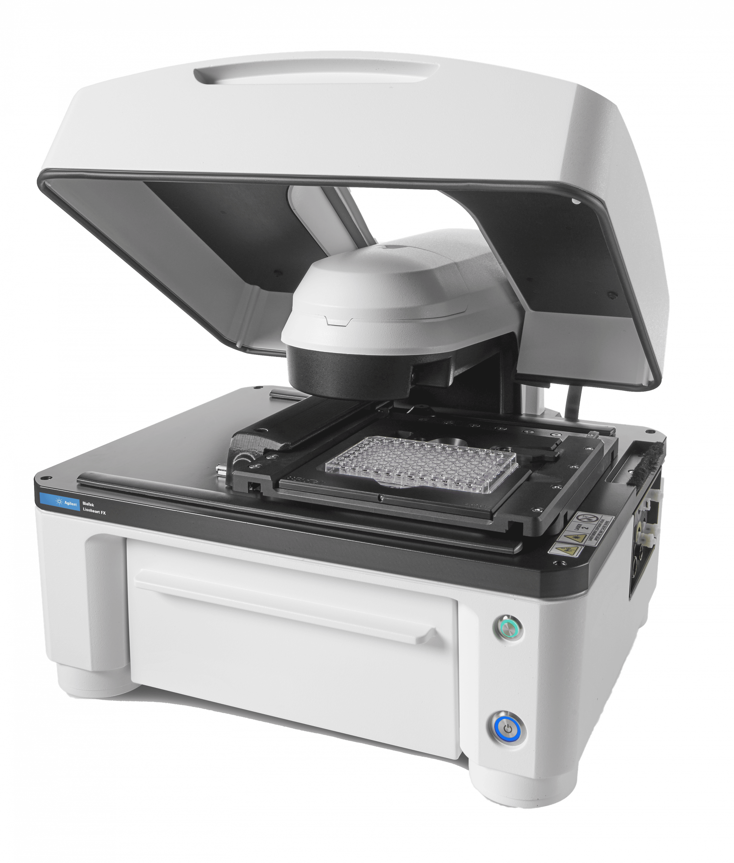
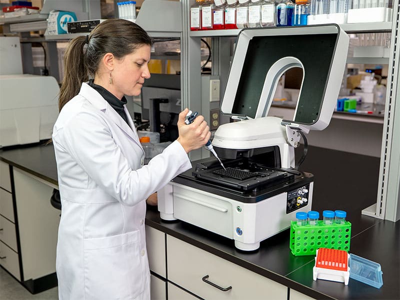
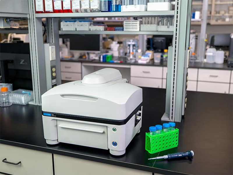
Lionheart™ FX Automated Microscope is a compact, inclusive microscopy system for a broad range of imaging workflows.
It offers up to 100x air and oil immersion magnification, with:
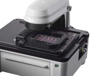 An optional environment control cover provides incubation to 40°C and effective containment for CO2/O2 control, a humidity chamber optimizes conditions for long-term live cell imaging applications, and an available dual reagent injector facilitates rapid kinetic assays. Lionheart FX and Gen5 together comprise Augmented Microscopy™ – the automation of image capture, processing, analysis and development of publication-ready images and data.
| Lionheart FX | Lionheart LX | |
| General | ||
| Microplate types | 6- to 1536-well plates | 6- to 1536-well plates |
| Other labware supported | Microscope slides, Petri and cell culture dishes, cell culture flasks (T25, T75), counting chambers (hemocytometers), chamber slides | Microscope slides, Petri and cell culture dishes, cell culture flasks (T25, T75), counting chambers (hemocytometers), chamber slides |
| Support for labware up to 1.5″ tall | Support for labware up to 1.5″ tall | |
| Temperature control | Incubation to 40 °C with optional environmental control cover | |
| Software | Gen5 Microplate Reader and Imager Software included | Gen5 Microplate Reader and Imager Software included |
| Gen5 Image and Image+ Software available for advanced image analysis (option) | Gen5 Image and Image+ Software available for advanced image analysis (option) | |
| CO2 and O2 control (option) | 0 – 20% CO2 control and 1 – 19% O2 control, with optional Gas Controller | |
| Environmental control cover | Top cover for light tight imaging and incubation control (option) | |
| X/Y Stage resolution | Lead screw driven stage with 0.1 micron resolution | Lead screw driven stage with 0.1 micron resolution |
| Humidity control | Humidity chamber with rapid gas recharge (option) | |
| Physical Characteristics | ||
| Power | 250 Watts max. | 250 Watts max. |
| Dimensions | With cover closed or without cover: 18.3″ D, 17.9″ W, 14.1″ H (46.5 cm x 45.5 cm x 35.8 cm) | 18.3″ D, 17.9″ W, 14.1″ H (46.5 cm x 45.5 cm x 35.8 cm) |
| With cover fully open: 18.3″ D, 17.9″ W, 27.5″ H (46.5 cm x 45.5 cm x 69.8 cm) | ||
| Weight | Without environmental control cover: 51 lbs (23.1 kg) | 51 lbs (23.1 kg) |
| With environmental control cover: 58 lbs (26.3 kg) | ||
| Regulatory | ||
| Regulatory | CE and TUV marked. RoHS compliant. Models for In Vitro Diagnostic use are available. | CE and TUV marked. RoHS compliant. Models for In Vitro Diagnostic use are available. |
| Imaging System | ||
| Imaging mode | Fluorescence, brightfield, color brightfield, and phase contrast | Fluorescence, high contrast brightfield and color brightfield |
| Imaging method | Single color, multi-color, montage, time lapse, Z-stacking, burst mode | Single color, multi-color, montage, time lapse, Z-stack, z-stack montage, burst mode |
| Light source | High power LEDs (available wavelengths: 365 nm, 390 nm, 465 nm, 505 nm, 523 nm, 590 nm, 623 nm, 655 nm, 740 nm) | High power LEDs (available wavelengths: 365 nm, 390 nm, 465 nm, 505 nm, 523 nm, |
| 590 nm, 623 nm, 655 nm, 740 nm) | ||
| Camera | 16-bit gray scale, Sony CCD, 1.25 megapixel | 16-bit CMOS camera. 2.3 MP Sony IMX249 1/1.2″ sensor with 5.86 micron pixel size |
| Camera binning | Optional 2×2 binning for focus and/or image capture | Optional 2×2 binning for focus and/or image capture |
| Camera exposure range | 5 milliseconds to 4 seconds | 5 milliseconds to 4 seconds |
| Image outputs available | Raw Images: 16-bit TIFF | Raw Images: 16-bit TIFF |
| Saved Images: TIF, JPG, BMP, PNG, EMF, GIF | Saved Images: TIFF, JPG, BMP, PNG, EMF, GIF, color TIFF | |
| Movies: MP4, WMV | Movies: MP4, WMV | |
| Objective capacity | 6 onboard, user-replaceable objectives | 6 onboard, user-replaceable objectives |
| Objectives available | Fluorescence: | Air: 1.25x, NA .04; 2.5x (2.25x eff), NA: .07; 2.5x (2.75x eff), NA: .12; 4x, NA: .13; 10x, NA: .30; 20x, NA: .45; 40x, NA.60; 60x, NA .70 |
| Air: 1.25x, NA .04; 2.5x (2.25x eff), NA: .07; 2.5x (2.75x eff), NA: .12; 4x, NA: .13; 10x, NA: .30; | Oil: 60x, NA:1.42; 60x, NA 1.25; 100x, NA 1.4; 100x, NA 1.3 | |
| 20x, NA: .45; 40x, NA.60; 60x, NA .70 | High NA: 20x, NA 0.75; 40x, NA 0.95 | |
| Oil: 60x, NA:1.42; 100x, NA: 1.40 | ||
| Phase objectives available: 4x, NA: .13; 10x, NA: .30; 20x, NA: .45; 40x, NA .60 | ||
| Image filter cube capacity | 4 user-replaceable fluorescence cubes plus brightfield channel | 4 user-replaceable fluorescence cubes plus brightfield channel |
| Imaging filter cubes available | DAPI, CFP, GFP, YFP, RFP, Texas Red, CY5, CY7, Acridine Orange, CFP-YFP FRET, Chlorophyll, Phycoerythrin (PE), Propidium Iodide, CY5.5, TagBFP, GFP (Ex)-CY6 (Em), RFP (Ex)-CY5 (Em), Alexa 568, Ex377 / Em647 | DAPI, CFP, GFP, YFP, RFP, Texas Red, CY5, CY7, Acridine Orange, CFP-YFP FRET, Chlorophyll, Phycoerythrin (PE), Propidium Iodide, CY5.5, TagBFP, GFP (Ex)-CY6 (Em), RFP (Ex)-CY5 (Em), Alexa 568, Ex377 / Em647 |
| Automated functions | Autofocus, user-trained autofocus, auto exposure, auto-LED intensity | Autofocus, user-trained autofocus, auto exposure, auto-LED intensity |
| Autofocus method | Image-based autofocus | Image-based autofocus |
| Laser autofocus option | Laser autofocus option | |
| Image collection rate | Single well fastest frame rate capture: | Single well fastest frame rate capture: |
| *Full Resolution: up to 10 frames per second for single color images | *Full Resolution: up to 10 frames per second for single color images | |
| *2×2 Binning: up to 20 frames per second for single color images | *2×2 Binning: up to 20 frames per second for single color images | |
| Microscope stage control | Gen5 Software Control | Gen5 Software Control |
| Optional joystick controller | Optional joystick controller | |
| Reagent Injectors (option) | ||
| Number | 2 syringe pumps | |
| Supported labware | 6- to 384-well microplates, Petri and cell culture dishes, chamber slides | |
| Dead volume | <1.65 mL with back flush | |
| Dispense tip options | Aligned tip – aligned with optical path for dispensing for fast kinetic assays | |
| Offset tip – dispensing is offset from the optical path | ||
| Dispense volume | 5 – 1000 µL in 1 µL increments | |
| Dispense accuracy | +1 µL or 2% | |
| Dispense precision | <2% at 50 – 200 µL | |
| Specifications are subject to change. |
| GENERAL | |
| Microplate types | 6- to 1536-well plates |
| Other labware supported | Microscope slides, Petri and cell culture dishes, cell culture flasks (T25, T75), counting chambers (hemocytometers), chamber slides |
| Support for labware up to 1.5″ tall | |
| Temperature control | Incubation to 40 °C with optional environmental control cover |
| Software | Gen5 Microplate Reader and Imager Software included |
| Gen5 Image and Image+ Software available for advanced image analysis (option) | |
| CO2 and O2 control (option) | 0 – 20% CO2 control and 1 – 19% O2 control, with optional Gas Controller |
| Environmental control cover | Top cover for light tight imaging and incubation control (option) |
| X/Y Stage resolution | Lead screw driven stage with 0.1 micron resolution |
| Humidity control | Humidity chamber with rapid gas recharge (option) |
| IMAGING SYSTEM | |
| Imaging mode | Fluorescence, brightfield, color brightfield, and phase contrast |
| Imaging method | Single color, multi-color, montage, time lapse, Z-stacking, burst mode |
| Light source | High power LEDs (available wavelengths: 365 nm, 390 nm, 465 nm, 505 nm, 523 nm, 590 nm, 623 nm, 655 nm, 740 nm) |
| Camera | 16-bit gray scale, Sony CCD, 1.25 megapixel |
| Camera binning | Optional 2×2 binning for focus and/or image capture |
| Camera exposure range | 5 milliseconds to 4 seconds |
| Image outputs available | Raw Images: 16-bit TIFF |
| Saved Images: TIF, JPG, BMP, PNG, EMF, GIF | |
| Movies: MP4, WMV | |
| Objective capacity | 6 onboard, user-replaceable objectives |
| Objectives available | Fluorescence: |
| Air: 1.25x, NA .04; 2.5x (2.25x eff), NA: .07; 2.5x (2.75x eff), NA: .12; 4x, NA: .13; 10x, NA: .30; | |
| 20x, NA: .45; 40x, NA.60; 60x, NA .70 | |
| Oil: 60x, NA:1.42; 100x, NA: 1.40 | |
| Phase objectives available: 4x, NA: .13; 10x, NA: .30; 20x, NA: .45; 40x, NA .60 | |
| Image filter cube capacity | 4 user-replaceable fluorescence cubes plus brightfield channel |
| Imaging filter cubes available | DAPI, CFP, GFP, YFP, RFP, Texas Red, CY5, CY7, Acridine Orange, CFP-YFP FRET, Chlorophyll, Phycoerythrin (PE), Propidium Iodide, CY5.5, TagBFP, GFP (Ex)-CY6 (Em), RFP (Ex)-CY5 (Em), Alexa 568, Ex377 / Em647 |
| Automated functions | Autofocus, user-trained autofocus, auto exposure, auto-LED intensity |
| Autofocus method | Image-based autofocus |
| Laser autofocus option | |
| Image collection rate | Single well fastest frame rate capture: |
| *Full Resolution: up to 10 frames per second for single color images | |
| *2×2 Binning: up to 20 frames per second for single color images | |
| Microscope stage control | Gen5 Software Control |
| Optional joystick controller | |
| REAGENT INJECTORS (OPTION) | |
| Number | 2 syringe pumps |
| Supported labware | 6- to 384-well microplates, Petri and cell culture dishes, chamber slides |
| Dead volume | <1.65 mL with back flush |
| Dispense tip options | Aligned tip – aligned with optical path for dispensing for fast kinetic assays |
| Offset tip – dispensing is offset from the optical path | |
| Dispense volume | 5 – 1000 µL in 1 µL increments |
| Dispense accuracy | +1 µL or 2% |
| Dispense precision | <2% at 50 – 200 µL |
| PHYSICAL CHARACTERISTICS | |
| Power | 250 Watts max. |
| Dimensions | With cover closed or without cover: 18.3″ D, 17.9″ W, 14.1″ H (46.5 cm x 45.5 cm x 35.8 cm) |
| With cover fully open: 18.3″ D, 17.9″ W, 27.5″ H (46.5 cm x 45.5 cm x 69.8 cm) | |
| Weight | Without environmental control cover: 51 lbs (23.1 kg) |
| With environmental control cover: 58 lbs (26.3 kg) | |
| REGULATORY | |
| Regulatory | CE and TUV marked. RoHS compliant. Models for In Vitro Diagnostic use are available. |
Register to receive email updates on our new products, special promotions and events.
Home » Instruments » BioTek Lionheart FX
© Copyright 2025. All Rights Reserved.
A Beun - De Ronde company