xCELLigence RTCA CardioECR
Combines high frequency measurement of cell-induced electrical impedance with multi-electrode array technology to simultaneously assess cardiomyocyte contractility, viability, and electrophysiology
Combines high frequency measurement of cell-induced electrical impedance with multi-electrode array technology
Unprecedented level of detail

Information

Cardiomyocyte Contractility & Electrical Activity

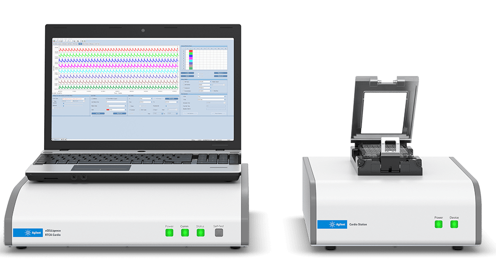
The xCELLigence RTCA CardioECR instrument combines high frequency measurement of cell-induced electrical impedance with multi-electrode array technology to simultaneously assess cardiomyocyte contractility, viability, and electrophysiology. The simultaneous recording of impedance and field potential by the xCELLigence RTCA CardioECR instrument provides a view of cardiomyocyte health at an unprecedented level of detail, enabling a deeper understanding of the mechanisms underlying drug-induced cardiac liability.

The CardioECR can also be used to functionally mature hiPSC cardiomyocytes with its electronic pacing function. Paced hiPSC cardiomyocytes provides a significantly improved cell models used in various applications including safety/tox assessment, drug discovery, and cardiac disease research.


Features:

  • Monitor excitation-contraction coupling and integrated ion channel activity
  • Generate data that highly correlates with arrhythmogenic activity
  • Use the pacing function for more tightly controlled assays
  • Obtain a more thorough understanding of drug mechanism of action


Cellular Impedance Explained

Positioned between reductionistic biochemical assays and whole organism in vivo experimentation, cell-based assays serve as an indispensable tool for basic and applied biological research. However, the utility of many cell-based assays is diminished by:

  1. The need to use labels
  2. Incompatibility with continuous monitoring (i.e. only end point data is produced),
  3. Incompatibility with orthogonal assays
  4. The inability to provide an objective/quantitative readout.

Each of these shortcomings is, however, overcome by the non-invasive, label-free, and real-time cellular impedance assay.

Find out more on the website of Agilent

Cardio CardioECR
Recording in incubator  


System
IMP electrodes  


FP electrodes  

 
Electrical pacing function  

 
device
plate 96-well E-Plate 48-well E-Plate
Application
Cell attachment and viability  


Contractility  


FP (ion channels) assessment  

 
Acute assay (seconds to minutes)  


Long-term assay (hours to days)  


recording Recording in incubator 96 wells, 12.9 ms Up to 24 wells -1ms; all 48 wells – 2ms
Field potential sampling rate n/a 10 kHz
Sampling capability Simultaneous recording of impedance in 96 wells Simultaneous recording of impedance and field potential electrodes in all 48 wells
stimulation Stimulation voltage range n/a -2.5 V to +2.5 V
Simultaneous stimulation n/a Up to 48 wells (across the entire plate)
 
Analyzer Application Apoptosis Cell Characterization Cytotoxicity
Proliferation Stem Cells Cardiotoxicity
Depth 34 cm
Height 16 cm
Operating Environment Relative Humidity 5-98 %
Operating Environment Temperature 15-40 °C
Sampling Format 96-well plate
Width 28 cm
Cardiac Disease Modeling The pathophysiological cellular phenotypes of genetically heritable heart diseases can be modeled using disease-specific human induced pluripotent stem cell cardiomyocyte (hiPSC-CM). Modeling enables broader understanding of the mechanisms leading to compromised electrical and contractile coupling.
 
Cardio Safety Toxicology Human induced pluripotent stem cell cardiomyocytes (hiPSC-CM) have been used to assess drug liability. The xCELLigence RTCA CardioECR system offers a unique multiplex detection method to probe and understand the underlying toxicity of compounds and pharmaceuticals.
 
Cardio Drug Discovery Human induced pluripotent stem cell cardiomyocytes (hiPSC-CM) offer an exciting new model system to test both the efficacy and toxicity of new therapeutic approaches, including inotropic compounds that serve to modulate the force of cardiomyocyte contractility.
 
The CiPA (Comprehensive In Vitro Proarrhythmia Assay) Initiative Driven by HESI, FDA and Safety Pharmacology Society, the CiPA initiative aims to evaluate potential modifications to current regulatory FDA guidelines for cardiac safety assessment. The xCELLigence RTCA CardioECR was selected as a core technology for validation and has participated in Phase I and II.
CardioECR Impedance The xCELLigence RTCA CardioECR instrument combines impedance recording with both multi-electrode array (MEA) technology and a pacing function. Cardiomyocytes are seeded in a 48-well electronic microplates (E-Plate CardioECR 48) that contains biosensors fused to the bottom of each well (Figures 1A-C). The enhanced impedance measurement rate (every 1 ms) of the xCELLigence RTCA CardioECR system provides extremely high temporal resolution for viewing subtleties of the cardiomyocyte contraction/relaxation continuum. In addition to this ability to monitor cell viability and contractile activity, additional biosensors in the well bottoms (Figure 1C) allow for extracellular field potential (FP) measurements at 10 kHz that can be performed in tandem with impedance recording.
Contracted vs Relaxed comparison Application of a low voltage (less than 20 mV) establishes an electric current between the electrodes, which is differentially modulated by the number of cells covering the electrodes, the morphology of those cells, and the strength of cell attachment. Because the cardiomyocyte contraction/relaxation cycle involves substantial changes in cell morphology and adhesion, it can be dynamically monitored using impedance (Figures 2A-B). The enhanced impedance measurement rate (every 1 ms) of the xCELLigence RTCA CardioECR system provides extremely high temporal resolution for viewing subtleties of the cardiomyocyte contraction/relaxation continuum. In addition to this ability to monitor cell viability and contractile activity, additional point electrodes in the well bottoms allow for extracellular field potential (FP) measurements at 10 kHz, which can be performed in tandem with impedance recording.
CardioECR Software The RTCA CardioECR Software enables facile experiment setup and execution along with powerful data analysis, while remaining efficient and intuitive. Measure impedance and field potential simulateanously in real time and view the data traces overlaid (see figure) or individually.
Cellular Impedance Explained Positioned between reductionistic biochemical assays and whole organism in vivo experimentation, cell-based assays serve as an indispensable tool for basic and applied biological research. However, the utility of many cell-based assays is diminished by: (1) the need to use labels, (2) incompatibility with continuous monitoring (i.e. only end point data is produced), (3) incompatibility with orthogonal assays, and (4) the inability to provide an objective/quantitative readout. Each of these shortcomings is, however, overcome by the non-invasive, label-free, and real-time cellular impedance assay.
Functional Unit of Cellular Impedance Assay The functional unit of a cellular impedance assay is a set of gold microelectrodes fused to the bottom surface of a microtiter plate well (Figure 1). When submersed in an electrically conductive solution (such as buffer or standard tissue culture medium), the application of an electric potential across these electrodes causes electrons to exit the negative terminal, pass through bulk solution, and then deposit onto the positive terminal to complete the circuit. Because this phenomenon is dependent upon the electrodes interacting with bulk solution, the presence of adherent cells at the electrode-solution interface impedes electron flow. The magnitude of this impedance is dependent on the number of cells, the size and shape of the cells, and the cell-substrate attachment quality. Importantly, neither the gold microelectrode surfaces nor the applied electric potential (22 mV) have an effect on cell health or behavior.
 
Impedance Electrodes The gold microelectrode biosensors in each well of ACEA’s electronic microtiter plates (E-Plates®) cover 70-80% of the surface area (depending if a view area is present). Rather than the simplified electrode pair depicted in Figure 1, the electrodes in each well of an E-Plate are linked into “strands” that form an interdigitating array (Figure 2). This arrangement enables populations of cells to be monitored simultaneously and thereby provides exquisite sensitivity to: the number of cells attached to the plate, the size/morphology of the cells, and the cell-substrate attachment quality.
Real-Time Impedance Traces Explained The impedance of electron flow caused by adherent cells is reported using a unitless parameter called Cell Index (CI), where CI = (impedance at time point n – impedance in the absence of cells)/nominal impedance value. Figure 3 provides a generic example of a real-time impedance trace throughout the course of setting up and running an apoptosis experiment. For the first few hours after cells have been added to a well there is a rapid increase in impedance. This is caused by cells falling out of suspension, depositing onto the electrodes, and forming focal adhesions. If the initial number of added cells is low and there is empty space on the well bottom cells will proliferate, causing a gradual yet steady increase in CI. When cells reach confluence the CI value plateaus, reflecting the fact that the electrode surface area that is accessible to bulk media is no longer changing. The addition of an apoptosis inducer at this point causes a decrease in CI back down to zero. This is the result of cells rounding and then detaching from the well bottom. While this generic example involves drug addition when cells are confluent, impedance-based assays are extremely flexible and can also evaluate the rate and extent of initial cell adhesion to the electrodes, or the rate and extent of cell proliferation.
 
Correlating Impedance with Cellular Phenomena RTCA provides a quantitative readout of cell number, proliferation rate, cell size/shape, and cell-substrate attachment quality. Because these physical properties are the product of thousands of different genes/proteins, RTCA can provide an extremely wide field of view on cell health and behavior. Everything from endothelial barrier function and chemotaxis to filopodia dynamics and immune cell-mediated cytolysis have successfully been analyzed on xCELLigence instruments. Despite the breadth of their reach, xCELLigence assays are still capable of interrogating very specific biochemical and cellular phenomena. Appropriate use of controls and/or orthogonal techniques make it possible to correlate the features of an impedance trace with specific cellular/molecular phenomena. To learn more about how this is done, and to witness the sensitivity and versatility of the xCELLigence RTCA technology, peruse the many specific applications that are highlighted here.
The xCELLigence RTCA CardioECR instrument combines high frequency measurement of multi-electrode array technology with cell-induced electrical impedance to simultaneously assess cardiomyocyte contractility, viability, and electrophysiology. The simultaneous recording of the field potential and impedance by the xCELLigence RTCA CardioECR instrument provides a view of cardiomyocyte health at an unprecedented level of detail. The enables a deeper understanding of the mechanisms underlying drug-induced cardiac liability. The CardioECR can also be used to functionally mature hiPSC cardiomyocytes with its electronic pacing function. Paced hiPSC cardiomyocytes provide a significantly improved cell models used in various applications including safety/tox assessment, drug discovery, and cardiac disease research. Key Features • Monitor excitation-contraction coupling and integrated ion channel activity • Generate data that highly correlates with arrhythmogenic activity • Use the pacing function for more tightly controlled assays • Obtain a more thorough understanding of drug mechanism of action Typical Applications • Cardio Maturation: Improve the maturation status of human induced pluripotent stem cell cardiomyocytes (hiPSC-CM). • Cardiac disease modelling: Studying the phenotypes of genetically heritable heart diseases with disease-specific human induced pluripotent stem cell cardiomyocytes (hiPSC-CM). • Cardio Safety Toxicology: Assess the drugs liability and underlying toxicity of medicine and pharmaceuticals. • Cardio Drug Discovery: Test the efficacy and toxicity of pharmaceuticals with the usage of human induced pluripotent stem cell cardiomyocytes (hiPSC-CM).

Related instruments

xcelligence-rtca-cardio
xCELLigence RTCA Cardio
Monitors cardiomyocyte activity for cardiotoxicity assessment
Monitor cardiomyocyte activity for cardiotoxicity assessment
Real-time control and monitoring
xcelligence mp
xCELLigence RTCA MP - Multiple plates
Obtain continuous cell assay data points using label-free methods, in a 6 x 96-well format
Run up to six 96-well plates simultaneously or independently
Easy workflow
Intuitive RTCA Software Pro
xcelligence-rtca-s16
xCELLigence RTCA S16 - Pilot scale
Continously monitor live cell proliferation, morphology change, and attachment quality in a label-free and real-time manner
Continously monitoring of live cell proliferation, morphology change, and attachment quality
Non-invasive and label-free real-time analysis

Recently viewed instruments

No links are found