xCELLigence RTCA SP
Single plate RTCA Analyzer with uses biosensors to monitor in real-time cell proliferation & viability, biofilm formation & dissipation, and cytophathic effects in a label-free manner
Non-invasive and label-free real-time impedance technology 
Simply add cells to E-plates and begin kinetic measurements
Minimal hands-on time  

Information

The Agilent xCELLigence Real-Time Cell Analysis (RTCA) SP instrument uses label-free cellular impedance to continuously monitor cell health, behavior, and function with high accuracy, sensitivity, and reproducibility. The SP model can run a 96-well electronic microplate (E-Plate 96), which is ideal for cell health characterization, immune cell-mediated killing, viral cytopathic effects, cytotoxicity, cell adhesion, cell barrier function (TEER), cell signaling (e.g., GPCR) and other related applications.

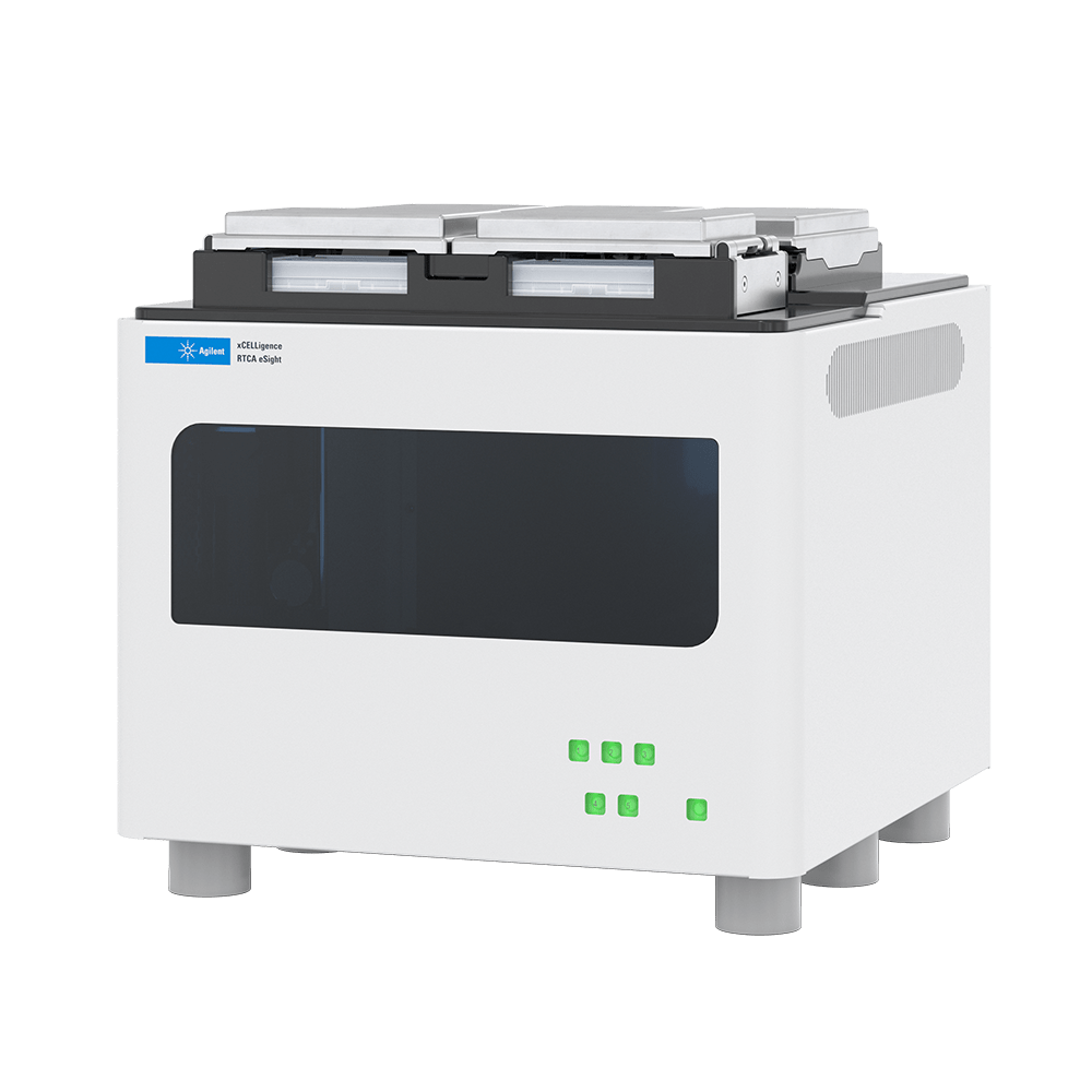
The instrument operates in a standard CO2 cell culture incubator and the control unit is housed outside the incubator. Simply plate cells and begin monitoring cell behavior to obtain real-time kinetic data for assay windows that stretch from seconds to days. Online data acquisition and offline data analysis are easy to perform using RTCA Software Pro, which also supports FDA 21 CFR Part 11 compliance.


Key features:

  • xCELLigence RTCA SP model can run one 96-well plate
  • Measure cell health, viability, or response to treatments with high sensitivity, accuracy, and reproducibility
  • Obtain continuous data at high temporal resolution (from seconds to days), with non-invasive and label-free real-time impedance technology
  • Easy workflow allows users to simply add cells to E-plates and begin kinetic measurements at physiological conditions, with minimal hands-on time
  • Intuitive RTCA Software Pro allows for analysis of real-time data to generate diverse plot types and automatically calculate parameters such as % of cytolysis, IC50 or KT50
  • RTCA Software Pro supports FDA 21 CFR Part 11 compliance to ensure electronic data integrity
Analyzer Application Adhesion Apoptosis Cell Characterization
Cytotoxity Immune Cell Killing Proliferation
Receptor Signaling Stem Cells Virus Cytopathic Effects
Wound Healing
Depth 12 cm
Height 12 cm
Operating Environment Relative Humidity 5-98 %
Operating Environment Temperature 20-40 °C
Sampling Format 96-well plate
Width 23 cm

Immune Cell Killing Assays

Typical immune cell killing assays are labor-intensive and require users to collect multiple timepoints using a combination of techniques. In contrast, the label-free xCELLigence RTCA instruments provide a complete view of immune cell killing and closely mimics activity in vivo. Obtain highly sensitive, reproducible, and direct measurements of immune cell killing potency in real time. This simple and high throughput workflow also provides greater insight into cellular mechanism of action, serial killing, and exhaustion.  

Viral Cytopathic Effect Assays

Advance your anti-viral therapy or vaccine development by accurately measuring viral cytopathic effects (CPE) in real time with Agilent xCELLigence RTCA instruments, without the use of labor-intensive plaque assays. Monitor viral CPE automatically, from minutes to days, and greatly reduce workload and manual handling of samples. Measure cell proliferation kinetics, identify the optimal time point for viral infection with different cell seeding densities, and assess virus-mediated cytopathogenicity in one workflow.  

Cytotoxicity Assays

Determine cytotoxic effects of drugs or compounds by monitoring cell proliferation, cell size or morphology, and cell-substrate attachment quality in real time. Monitor the kinetics of long-term cellular responses with this simple and high-throughput cytotoxicity assay. Make informed decisions about the timing of cell treatments and easily generate dose-response curves at multiple time points, without increasing the workload.  

Cell Adhesion Assays

During tumor formation, primary cancers shed millions of cells into circulation, which are known as circulating tumor cells (CTCs). CTCs can re-attach, extravasate, and metastasize in distant organs.  With xCELLigence RTCA, continuously monitor cell adhesion kinetics of different cell types to gain deeper insight into the intricacy of metastasis. Quantitatively assess cell attachment and spreading in real time, without the use of fluorescent reagents or dyes.  

Cell Signaling Assays

G protein-coupled receptor (GPCR), receptor tyrosine kinase (RTK), or nuclear hormone-mediated signaling often results in biochemical changes that affect the number of cells present, the size/morphology of the cells, and how tightly the cells are interacting with the plate surface. With xCELLigence RTCA, you can sensitively capture changes in cell size, shape, and proliferation rate in response to cell signaling events. This label-free and continuous monitoring provides quantitative assessment of cell response in the initial minutes of exposure and their response over days.  

Stem Cell Assays

Stem cell research provides mechanistic insights into disease, aids regenerative medicine development, and offers an alternative approach to test the liability and effectiveness of new drugs. Overcome cell therapy manufacturing challenges by predicting functional capacity and assessing variability of mesenchymal stem cells (MSCs) with xCELLigence RTCA. Maximize production yields using fewer cells and continuously measure integrated changes in cell number, attachment, and morphology to ensure consistency between passages.  

Cell Barrier Function Assays

Cell barrier function, provided by endothelial and epithelial cells, can become compromised in many disease states. Endothelial cells line the interior surface of blood vessels and lymphatic vessels. Epithelial cells form the surfaces of the body that serve as a barrier to protect from foreign molecules. The impedance-based xCELLigence RTCA assay is a sensitive and quantitative alternative to the traditional methods to measure barrier disruption, including the solute permeability assay and transendothelial electrical resistance (TEER) assay.
Cellular Impedance Explained Positioned between reductionistic biochemical assays and whole organism in vivo experimentation, cell-based assays serve as an indispensable tool for basic and applied biological research. However, the utility of many cell-based assays is diminished by: (1) the need to use labels, (2) incompatibility with continuous monitoring (i.e. only end point data is produced), (3) incompatibility with orthogonal assays, and (4) the inability to provide an objective/quantitative readout. Each of these shortcomings is, however, overcome by the non-invasive, label-free, and real-time cellular impedance assay.
Functional Unit of Cellular Impedance Assay The functional unit of a cellular impedance assay is a set of gold microelectrodes fused to the bottom surface of a microtiter plate well (Figure 1). When submersed in an electrically conductive solution (such as buffer or standard tissue culture medium), the application of an electric potential across these electrodes causes electrons to exit the negative terminal, pass through bulk solution, and then deposit onto the positive terminal to complete the circuit. Because this phenomenon is dependent upon the electrodes interacting with bulk solution, the presence of adherent cells at the electrode-solution interface impedes electron flow. The magnitude of this impedance is dependent on the number of cells, the size and shape of the cells, and the cell-substrate attachment quality. Importantly, neither the gold microelectrode surfaces nor the applied electric potential (22 mV) have an effect on cell health or behavior.
 
Real-Time Impedance Traces Explained The impedance of electron flow caused by adherent cells is reported using a unitless parameter called Cell Index (CI), where CI = (impedance at time point n – impedance in the absence of cells)/nominal impedance value. Figure 3 provides a generic example of a real-time impedance trace throughout the course of setting up and running an apoptosis experiment. For the first few hours after cells have been added to a well there is a rapid increase in impedance. This is caused by cells falling out of suspension, depositing onto the electrodes, and forming focal adhesions. If the initial number of added cells is low and there is empty space on the well bottom cells will proliferate, causing a gradual yet steady increase in CI. When cells reach confluence the CI value plateaus, reflecting the fact that the electrode surface area that is accessible to bulk media is no longer changing. The addition of an apoptosis inducer at this point causes a decrease in CI back down to zero. This is the result of cells rounding and then detaching from the well bottom. While this generic example involves drug addition when cells are confluent, impedance-based assays are extremely flexible and can also evaluate the rate and extent of initial cell adhesion to the electrodes, or the rate and extent of cell proliferation.
Correlating Impedance with Cellular Phenomena RTCA provides a quantitative readout of cell number, proliferation rate, cell size/shape, and cell-substrate attachment quality. Because these physical properties are the product of thousands of different genes/proteins, RTCA can provide an extremely wide field of view on cell health and behavior. Everything from endothelial barrier function and chemotaxis to filopodia dynamics and immune cell-mediated cytolysis have successfully been analyzed on xCELLigence instruments. Despite the breadth of their reach, xCELLigence assays are still capable of interrogating very specific biochemical and cellular phenomena. Appropriate use of controls and/or orthogonal techniques make it possible to correlate the features of an impedance trace with specific cellular/molecular phenomena. To learn more about how this is done, and to witness the sensitivity and versatility of the xCELLigence RTCA technology, peruse the many specific applications that are highlighted here.

Related instruments

xCELLigence_eSight_web
xCELLigence RTCA eSight
Combining label-free impedance technology with brightfield imaging
Whole well imaging expansion
3 fluorescent channels spanning
Single setup for dual measurements
xcelligence mp
xCELLigence RTCA MP - Multiple plates
Obtain continuous cell assay data points using label-free methods, in a 6 x 96-well format
Run up to six 96-well plates simultaneously or independently
Easy workflow
Intuitive RTCA Software Pro

Recently viewed instruments

No links are found