| GENERAL |
|
| Detection modes |
UV-Vis absorbance |
| Fluorescence intensity |
| Luminescence |
| Fluorescence polarization |
| Time-resolved fluorescence |
| Alpha |
| Read methods |
Endpoint, kinetic, spectral scanning, well area scanning |
| Microplate types |
Monochromator: 6- to 384-well plates |
| Filters: 6- to 1536-well plates |
| Imaging: 6- to 1536-well plates |
| Other labware supported |
Microscope slides, Petri and cell culture dishes, cell culture flasks (T25), counting chambers (hemocytometer) |
| Take3 Micro-Volume Plates |
| Temperature control |
4-Zone incubation to 65 °C with Condensation Control |
| +0.2 °C at 37 °C |
| Shaking |
Linear, orbital, double orbital |
| Software |
Gen5™ Microplate Reader and Imager Software |
| Gen5 Image+ and Image Prime software available for full image analysis |
| Gen5 Secure for 21 CFR Part 11 compliance (option) |
| Automation |
BioStack and 3rd party automation compatible |
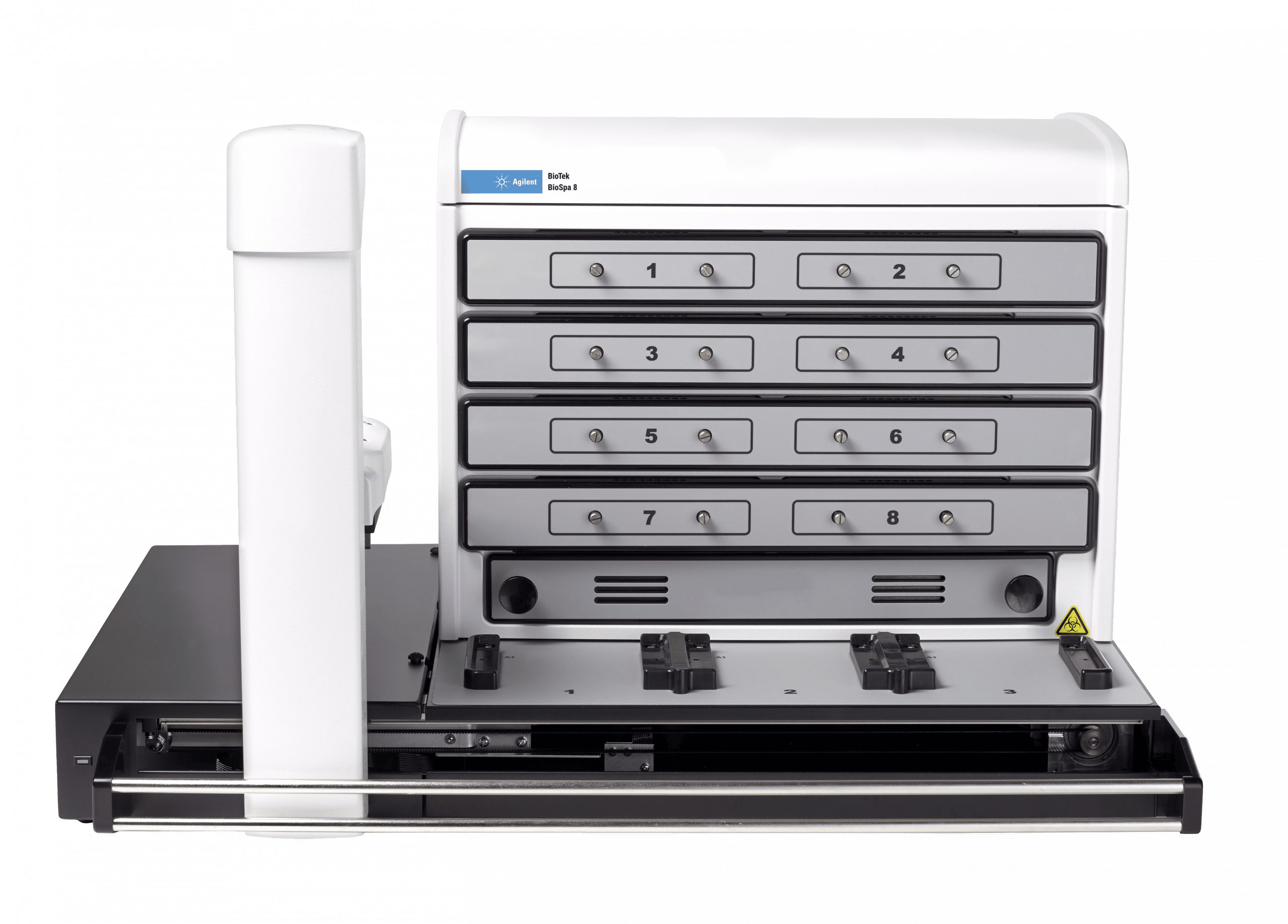
| BioSpa 8 Automated Incubator compatible |
| CO2 and O2 control (option) |
Range: 0 – 20% (CO2); 1 – 19% (O2) |
| Models for both CO2 and O2 or CO2 only are available |
| IMAGING SYSTEM |
|
| Imaging mode |
Fluorescence |
| Brightfield |
| Color Brightfield |
| Phase Contrast |
| Imaging method |
Single color, multi-color, montage, time lapse, z-stacking |
| Image processing |
Z-projection, digital phase contrast, stitching |
| Camera |
16-bit gray scale, Sony CCD |
| Objective capacity |
6 user-replaceable objectives |
| Objectives available |
1.25x, 2.5x (2.25x eff),2.5x (2.75x eff), 4x,10x, 20x, 40x, 60x |
| Phase objectives available |
4x, 10x, 20x, 40x |
| Image filter cube capacity |
4 user-replaceable fluorescence cubes plus brightfield channel |
| Imaging filter cubes available |
DAPI, CFP, GFP, YFP, RFP, Texas Red, CY5, CY7,Acridine Orange (ACR OR), CFP-YFP FRET, propidium,Iodide, chlorophyll, phycoerythrin, CY5.5, TagBFP, Alexa568, Ex377 / Em647 |
| Imaging LED cubes available |
365 nm, 390 nm, 465 nm, 505 nm, 523 nm, 590 nm, 623 nm, 655 nm, 740 nm |
| Automated functions |
Autofocus, auto LED intensity, auto exposure |
| Autofocus method |
Image-based autofocus |
| User-trained autofocus |
| Laser autofocus (option) |
| Positional controls |
Software control |
| Joystick control (option) |
| Image collection rate |
Image-based autofocus: |
| 96 wells, 1 color (DAPI), 4x, 6 minutes |
| 96 wells, 3 colors, 4x, 12 minutes |
|
| Laser autofocus: |
| 96 wells, 1 color (DAPI), 4x, <3 minutes |
| 96 wells, 3 colors, 4x, <7 minutes, 30 seconds |
| Burst Mode: 10 fps, single well, single color at <= 5Oms integration time |
| Image Analysis Software option |
Gen5 Image+: Advanced image analysis |
| Gen5 Image Prime: Advanced image analysis |
| Gen5 Secure Image+: Advanced image analysis, 21 CFR Part 11 features |
| FLUORESCENCE INTENSITY |
|
| Light source |
Xenon flash |
| Detector |
PMT for monochromator system |
| PMT for filter system |
| Wavelength selection |
Quad monochromators (top/bottom) |
| Filters (top) |
| Wavelength range |
Monochromators: 250 – 700 nm (850 nm option) |
| Filters: 200 – 700 nm (850 nm option) |
| Monochromator bandwidth |
Variable, from 9 nm to 50 nm in 1 nm increments |
| Dynamic range |
7 decades |
| Sensitivity |
Filters: |
| Fluorescein 0.25 pM (0.025 fmol/well, 384-well plate) |
|
| Quad Monochromator: |
| Fluorescein 2.5 pM (0.25 fmol/well, 384-well plate) – top |
| Fluorescein 4 pM (0.4 fmol/well, 384-well plate) – bottom |
| Reading speed (kinetic) |
96 wells: 11 seconds |
| 384 wells: 22 seconds |
| LUMINESCENCE |
|
| Wavelength range |
300 – 700 nm |
| Dynamic range |
>6 decades |
| Sensitivity |
Monos: 20 amol ATP (flash) |
| Filters: 10 amol ATP (flash), 100 amol (glow) |
| FLUORESCENCE POLARIZATION |
|
| Light source |
Xenon flash |
| Detector |
PMT |
| Wavelength selection |
Filters |
| Wavelength range |
280 – 700 nm (850 nm option) |
| Sensitivity |
1.2 mP standard deviation at 1 nM fluorescein |
| TIME-RESOLVED FLUORESCENCE |
|
| Light source |
Xenon flash |
| Detector |
PMT |
| Wavelength selection |
Quad monochromators (secondary mode) |
| Filters (top) |
| Wavelength range |
Filters: 200 – 700 nm (850 nm option) |
| Sensitivity |
Filters: Europium 40 fM (4 amol/well, 384-well plate) |
| Monos: Europium 1200 fM (120 amol/well, 384-well plate) |
| ABSORBANCE |
|
| Light source |
Xenon flash |
| Detector |
photodiode |
| Wavelength selection |
Monochromator |
| Wavelength range |
230 – 999 nm, 1 nm increment |
| Monochromator bandwidth |
4 nm (230 – 285 nm), 8 nm (>285 nm) |
| Dynamic range |
0 – 4.0 OD |
| Resolution |
0.0001 OD |
| Pathlength correction |
yes |
| Monochromator wavelength accuracy |
+2 nm |
| Monochromator wavelength repeatability |
+0.2 nm |
| OD accuracy |
<1% at 2.0 OD |
| <3% at 3.0 OD |
| OD linearity |
<1% from 0 to 3.0 OD |
| OD repeatability |
<0.5% at 2.0 OD |
| Stray light |
0.03% at 230 nm |
| Reading speed (kinetic) |
96 wells: 11 seconds |
| 384 wells: 22 seconds |
| ALPHA DETECTION |
|
| Light source |
100 mW 680 nm laser |
| Detector |
PMT |
| Wavelength selection |
Filters (top) |
| Sensitivity |
100 amol LCK peptide (384-well low volume plate) |
| REAGENT INJECTORS (OPTION) |
|
| Supported detection modes |
All modes |
| Number |
2 syringe pumps |
| Supported labware |
6- to 384-well plates, Petri and cell culture dishes |
| Dead volume |
1.1 mL with back flush |
| Dispense volume |
5 – 1000 µL in 1 µL increments |
| Plate geometry |
6- to 384-well microplates, Petri dishes |
| Dispense accuracy |
+1 µL or 2% |
| Dispense precision |
<2% at 50 – 200 µL |
| PHYSICAL CHARACTERISTICS |
|
| Power |
250 Watts max. |
| Dimensions |
20″ D x 16.5″ W x 17.5″ H |
| (50.8 cm x 41.91 cm x 44.5 cm) |
| Weight |
80 lbs (36.3 Kg) |
| REGULATORY |
|
| Regulatory |
CE and TUV marked. RoHS Compliant. Models for In Vitro Diagnostic use are available. |