BioTek Cytation C10
Confocal and widefield Imaging Reader. Combines spinning disk confocal and automated digital widefield microscopy, plus a multi-mode microplate reader.
Water-immersion objectives to capture more light with lower exposure times
Benchtop confocal imaging for every laboratory
Facilitate live cell imaging with environmental controls

Information

The Cytation C10 confocal imaging reader combines automated confocal and widefield microscopy with conventional multimode microplate reading in a unique, proprietary design. The automated water-immersion objectives capture more light, driving lower exposure times and reducing phototoxic impacts on live cells. The spinning-disk confocal module and accompanying disk pinhole options allow a researcher to look deeper into thick sample biology with improved clarity and detail. High-quality components used in the system, including a Hamamatsu scientific CMOS (sCMOS) camera, Olympus objectives and laser-based illumination enable excellent image quality, at a significantly lower price than others. Cytation C10 also includes widefield fluorescence, brightfield, and phase contrast optics.


Cytation C10 includes widefield fluorescence, brightfield, and phase contrast optics.

Onboard environmental controls enable live cell assays. Multiplate live cell assays are accomplished with Cytation C10 integrated with the Agilent BioTek BioSpa 8 automated incubator. Cytation C10 brings high-performance, affordable confocal to every laboratory.

  • Cytation C10 offers affordable, benchtop confocal imaging for every laboratory
  • Confocal microscopy enables improved image quality and analysis, revealing a level of detail in samples that is not possible with widefield optics
  • High-quality optical components allow the capture of stunning, publication-quality images
  • Water-immersion objectives to capture more light with lower exposure times
  • Combination of spinning disk confocal and widefield imaging plus multimode reader, allows the Cytation C10 to be ready for any assay with a modular, upgradable design
  • An available 60 µm deep sectioning disk (DSD) allows a clear view into thicker samples
  • Widefield imaging allows for faster acquisition of large samples at lower magnification, while confocal images small intracellular details or 3D samples, and both modes together provide highly multiplexed, multiparameter imaging experiments
  • Environmental controls, including incubation and CO2/O2 control and monitoring, facilitate live cell imaging
  • Imaging plus multimode detection and the hit-picking function quickly prescreen the microplate with the plate reader optics, then automatically image the samples that meet your hit criteria, saving time and data storage capacity
  • Variable bandwidth, quad monochromator plate reader optics enhance assay performance, including increased sensitivity and specificity
  • Combination of confocal and widefield imaging with multimode detection increases productivity with the ability to be ready for any application


Variety of applications from endpoint measurements to live cell imaging:

  • Cell viability/toxicity assays
  • 3D Cell Culture Assays
  • Cell Migration and Invasion Assays
  • Cell Proliferation
  • Label-free cell counting for cell proliferation studies
  • Cell Metabolism imaging and normalization – Seahorse data
  • Genotoxicity
  • Plaque assay
  • Fluorescent Microneutralization Assay


Configurations

Part # C10PHC2 C10MPHC2 C10PWC C10MPWC C10PW C10MPW
Confocal 60 µm spinning disk
Confocal 40 µm spinning disk
Hamamatsu sCMOS camera
Sony CMOS camera
Multi-mode reading
Phase contrast

Confocal: Improved image quality and analysis

Confocal microscopy can enable you to see a level of detail in your samples that is not possible with widefield optics. Not only can you obtain improved image quality, you can also get improved quantification and analysis with confocal images and Gen5 software.

GENERAL
Detection modes UV-Vis absorbance
Fluorescence intensity
Luminescence
Read methods Endpoint, kinetic, spectral scanning, well area scanning
Microplate types Monochromator: 6- to 384-well plates
Imaging: 6- to 1536-well plates
Other labware supported Microscope slides, Petri and cell culture dishes, cell culture flasks (T25), counting chambers (hemocytometer), Take3 Micro-volume plates
Temperature control 4-Zone incubation to 45 °C with Condensation Control
Shaking Linear, orbital, double-orbital
Software Gen5 Microplate Reader and Imager Software included
Gen5 Secure for 21 CFR Part 11 compliance (option)
Gen5 Image+ and Image Prime software available for full image analysis (option)
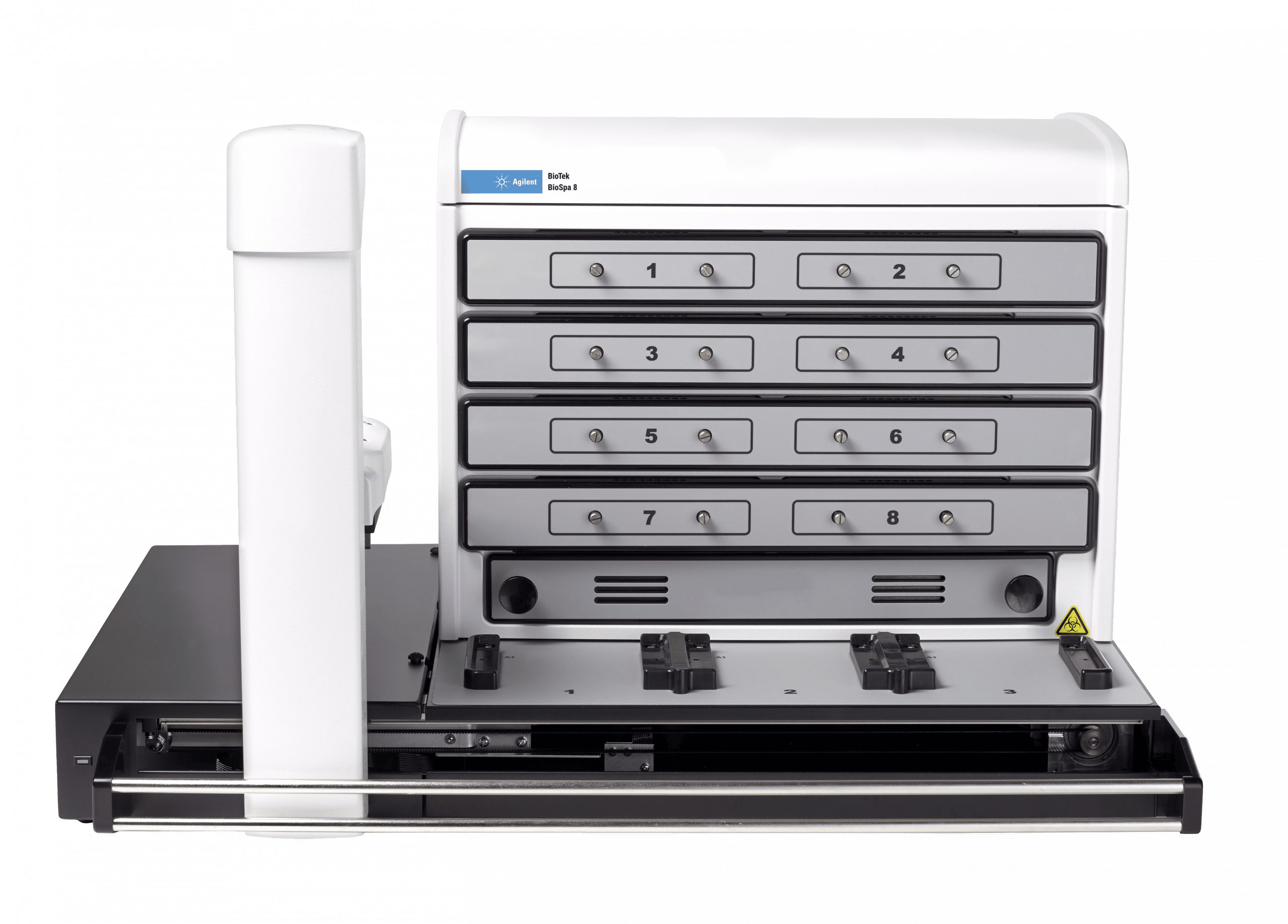
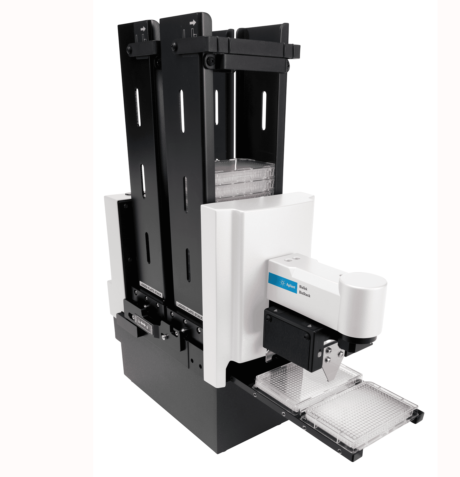
Automation BioStack and 3rd party automation compatible
CO2 and O2 control (option) Range: 0 – 20% (CO2); 1 – 19% (O2), with optional Gas Controller
Models for both CO2 and O2 or CO2 only are available
IMAGING – CONFOCAL MICROSCOPE
Imaging modes Fluorescence
Image processing Z-projection, digital phase contrast, stitching
Camera Hamamatsu Orca sCMOS, 16-bit grayscale camera or
Sony CMOS 16-bit grayscale camera
Objective capacity 6-position automated turret for user-replaceable objectives
Objectives available 20x, 40x, 60x
Image filter cube capacity 4 user-replaceable fluorescence cubes
Imaging filter cubes available CFP, CY5, DAPI, GFP, RFP, TRITC, brightfield
Laser 6-line
Automated functions Autofocus, user-trained autofocus, autoexposure, auto-LED intensity
Autofocus method Image-based autofocus
User-trained autofocus
Laser autofocus (option)
Positional controls Software Control
Joystick controller (option)
Imaging methods Single color, multi-color, time lapse, montage, z-stacking, z-stack montage
Field of view Hamamatsu sCMOS: 0.65 mm + 5% at 20x magnification
Sony CMOS: 0.70 mm + 5% at 20x magnification
Image collection rate Laser autofocus, 0 ms delay, 96 wells: 8 mins, 9 secs
Software autofocus, 0 ms delay, 96 wells: 12 mins, 1 sec
IMAGING – WIDEFIELD MICROSCOPE
Imaging mode Fluorescence, phase contrast, color brightfield, user-selectable brightfield/high contrast brightfield
Imaging method Single color, multi-color, time lapse, montage, z-stacking, z-stack montage
Image processing Z-projection, digital phase contrast, stitching
Camera Hamamatsu Orca sCMOS, 16-bit grayscale camera or
Sony CMOS 16-bit grayscale camera
Objective capacity 6-position automated turret for user-replaceable objectives
Objectives available 1.25x, 2.5x (2.25x eff), 4x,10x, 20x, 40x, 60x
Phase objectives available 4x, 10x, 20x, 40x
Image filter cube capacity 4 user-replaceable fluorescence cubes, plus brightfield channel
Imaging filter cubes available DAPI, CFP, GFP, YFP, RFP, Texas Red, CY5, CY7, Acridine Orange, CFP-FRET, CFP-YFP FRET, Chlorophyll, Phycoerythrin (PE), Propidium Iodide, CY5.5, TagBFP, Tag BFP-FRET, GFP (Ex)-CY5 (Em), RFP (Ex)-CY5 (Em), Alexa 568, Ex 377/Em 647, Oxidized roGFP2, TRITC
Imaging LED cubes available 365 nm, 390 nm, 465 nm, 505 nm, 523 nm, 554 nm, 590 nm, 623 nm, 655 nm, 740 nm
Automated functions Autofocus, autoexposure, auto-LED intensity
Autofocus method Image-based autofocus
User-trained autofocus
Laser autofocus (option)
Positional controls Software Control
Joystick controller (option)
Image collection rate Image-based autofocus:
96 wells, 1 color (DAPI), 4x, 6 minutes
Laser autofocus:
96 wells, 1 color (DAPI), 4x, <3 minutes
Image Analysis Software option Gen5 Image+: Image analysis
Gen5 Image Prime: Advanced image analysis
Gen5 Secure: 21 CFR Part 11 compliant features
FLUORESCENCE INTENSITY
Light source Xenon flash
Detector PMT
Wavelength selection Quad monochromators (top/bottom)
Wavelength range 250 – 700 nm (900 nm option)
Monochromator bandwidth Variable, from 9 nm to 50 nm in 1 nm increments
Dynamic range 7 decades
Reading speed (kinetic) 96 wells, sweep mode: 10 seconds
384 wells, sweep mode: 20 second
LUMINESCENCE
Wavelength range 300 – 700 nm
Dynamic range >6 decades
ABSORBANCE
Light source Xenon flash
Detector Photodiode
Wavelength selection Monochromator
Wavelength range 230 – 999 nm, 1 nm increments
Monochromator bandwidth 4 nm (230 – 285 nm), 8 nm (>285 nm)
Dynamic range 0 – 4.0 OD
Resolution 0.0001 OD
Pathlength correction Yes
Monochromator wavelength accuracy + 2 nm
Monochromator wavelength repeatability + 0.2 nm
OD accuracy <1% at 3.0 OD
OD linearity <1% from 0 to 3.0 OD
OD repeatability <0.5% at 2.0 OD
Stray light 0.03% at 230 nm
Reading speed (kinetic) 96 wells: 10 seconds
384 wells: 20 seconds
REAGENT INJECTORS (OPTION)
Supported detection modes All modes
Number 2 syringe pumps
Supported labware 6- to 384-well plates, Petri and cell culture dishes
Dead volume 1.1 mL, with back flush
Dispense volume 5 – 1000 µL in 1 µL increments
Plate geometry 6- to 384-well microplates
Dispense accuracy +1 µL or 2%
Dispense precision < 2% at 50 – 200 µL
PHYSICAL CHARACTERISTICS
Power Instrument: External 250W (minimum), 24VDC power supply, compatible with 100-240 VAC @50-60Hz.
Optional 6-channel laser light source: External 250W power supply, compatible with 100- 240VAC @ 50-60Hz.
Optional Hamamatsu scientific camera: External 75W power supply, compatible with 100- 240VAC @ 50-60Hz.
Dimensions 27″ W x 18.5″ H x 20″ D
(68.6 cm x 45.72 cm x 50.8 cm)
Weight 122 lbs (53.3 Kg)
REGULATORY
Regulatory CE and TUV marked. RoHS Compliant. Models for In Vitro Diagnostic use are available.
Gas Controller for CO2 and O2 controll Increasingly, life science assays are run on live cells. Some of these assays require control over CO2 and O2 concentrations to modulate the environment for pH buffering or to create a hypoxic condition. These assays are traditionally run-in flasks in a low-throughput manner, but there is a burgeoning interest in automation of live cell-based assays in microplates for higher throughput and efficiency, therefore there is a need for cell-friendly microplate instrumentation. The Gas Controller allows full control over CO2 and O2 concentrations to regulate the environment for microplate based live cell assays.
From whole organism imaging to high magnification subcellular image analysis, Gen5 helps transform raw data and images into meaningful results. Gen5 Software automates image capture from Agilent’s Lionheart Automated Microscope and Cytation Cell Imaging Multi-Mode Reader, and automates image processing and analysis to produce publication-ready images and quantitative data. This process of Augmented Microscopy™ enables simple, straightforward imaging workflows. • Automated image capture • Exceptional image processing tools • Powerful image analysis • Publication-ready images, graphs and data

Features

 
Augmented Microscopy automates image capture for samples with powerful tools for endpoint and time lapse workflows. Fluorescence, brightfield, color brightfield and phase contrast images are captured in slides, microplates, chamber slides, cell culture dishes for quantitative and qualitative analysis.
Exceptional Image Processing Tools Gen5’s image processing tools provide exceptional processing capability to facilitate the analysis of complex biologies, including 3D samples, large montaged samples and processes including live cell kinetics. From easy brightness/ contrast adjustments to deconvolution and automated movie file creation, image processing in Gen5 enables optimized visualization and analysis for many applications. The independent optical paths ensure uncompromised performance in all modes
Powerful Image Analysis Image analysis tools in Gen5 cover a very broad range of application requirements, and are both powerful and easy to use. Analysis functions in Gen5 extend to quantitative data as well. Cell count, confluence, signal translocation, subpopulation analysis and kinetic analysis are just some of the powerful data provided by Gen5’s image analysis tools.
Publication-ready Images, Graphs and Data Augmented Microscopy tools include the ability to create publication-ready images, graphs and data using the functions in Gen5 software. There is no need to export images or data to external software to create scatterplots, histograms, EC50 graphs and other data displays for publication.
Combined confocal and widefield microscopy Image quality is considerably improved due to the combination of spinning disk confocal and widefield microscopy. The degree of detail is exceptionally enhanced, and this results in improved quantification and analysis of the assay, combined with the Gen5 imaging Software.   High quality optical components To enable high quality images and publication-ready images, high standard objectives and filters are compatible with the Cytation C10. These high quality objectives and filters include the Hamamatsu sCMOS Orca, Semrock filters, Olympus objectives and other known brands.   Hit-picking: Multi-mode detection + imaging saves time and data storage The plate reader screens all wells and identifies the wells that meet certain criteria. Only the wells that meet these, will be imaged by the microscope. This results in the saving of time and computer memory! (1) Plate reader quickly identifies GFP positive wells. (2) Only GFP positive wells are imaged, saving both time and computer memory.Variable bandwidth monochromator for sensitivity and specificity The bandwidths of the monochromators in the Cytation C10 fluorescence detection module is selectable from 9 nm to 50 nm, in 1 nm increments. Variable bandwidths allow you to optimize detection conditions for fluorophores that have unique excitation or emission parameters, for example, a narrow Stokes shift. Adjusting the bandwidth results in better performance than conventional monochromators. The plate reader optics of Cytation use a quad monochromator design with variable bandwidth. The bandwidth can be set anywhere between 9 and 50 nm in 1 nm increment. Large bandwidth settings (1) provide increased sensitivity and lower limits of detection. Small bandwidth settings (2) provide increased specificity when multiple signals are present, which reduces signal crosstalk and enhances assay performanceMaintaining a consistent environment through CO2/O2 and temperature control Cytation C10 is well suited for live cell assays, with temperature control to 45 °C, plus linear and orbital shaking. A gas controller module is available to help regulate CO2 and O2 levels in the system for longer-term live cell requirements. A dual reagent injector provides quick inject/image capability for observing fast reactions. Successful live cell kinetic imaging relies on a consistent environment, including temperature control and CO2/O2 control and monitoring. Cytation C10 provides the perfect environment to grow and analyze live cells over time. Powerful movie maker and kinetic analysis software tools allow visualizing and analysis time-lapse experiments.Label-free cell counting It’s fast and convenient to count live cells using the available high contrast brightfield option. Higher contrast allows a direct and label-free method for object identification using Gen5 software to count cells and characterize cell proliferation in kinetic studies.

Related instruments

biospa_front-agilent
BioTek BioSpa 8
Automated Incubator
biostack4_square
BioTek BioStack 4
Compact and versatile microplate stacker
neo_center_closed-r-agilent
BioTek Synergy Neo2
Hybrid Multi-Mode Reader for demanding applications

Recently viewed instruments

No links are found